
The mTOR pathway is defined as a master regulatory kinase network that integrates nutrient availability, growth factor signals, energy status, and cellular stress to coordinate anabolic and catabolic processes. The role of mTOR pathway peptide research has expanded rapidly, with scientists now using purpose-designed peptides to probe both mTORC1 and mTORC2 complexes at the molecular level. mTORC1 drives protein and lipid synthesis while suppressing autophagy; mTORC2 governs cytoskeletal organization and Akt phosphorylation. Together, these complexes sit at the intersection of cancer, metabolic disease, and aging biology, making peptide signaling research in this space one of the most consequential areas in translational science.
How do peptides interact with and modulate the mTOR pathway?
Peptides modulate mTOR signaling through three distinct mechanisms: direct disruption of protein complexes, upstream anabolic activation, and engagement of negative regulators such as AMPK. Understanding which mechanism a peptide engages determines its research utility and therapeutic profile.
Direct complex disruption
Peptides derived from the mTOR-RHEB binding interface can competitively inhibit mTORC1 assembly. A 2026 study demonstrated that mutated interface peptides achieved binding energies of -14.53 kcal/mol compared to -15.83 kcal/mol for the native mTOR-RHEB complex. That near-native affinity confirms competitive inhibition is structurally achievable without full-length protein constructs. Hydrogen bonding patterns and intermolecular contact maps from HADDOCK 2.4 docking runs validate the binding geometry.

Anabolic activation via the GH-IGF-1 axis
Peptides that stimulate the growth hormone and IGF-1 axis activate mTORC1 to drive protein synthesis. GH secretagogues such as Sermorelin and Ipamorelin act upstream, triggering a cascade through PI3K, Akt, and TSC1/2 before reaching mTORC1. IGF-1 LR3 engages nutrient signaling receptors directly, producing a more sustained mTOR activation profile than endogenous IGF-1. These anabolic peptides are the primary tools for researchers studying protein synthesis, muscle hypertrophy models, and metabolic anabolism.
AMPK-mediated inhibition
A second peptide class activates AMPK, which phosphorylates TSC2 and Raptor to suppress mTORC1. The mitochondrial-derived peptide MOTS-c is the clearest example. MOTS-c activates AMPK, thereby inhibiting mTORC1 and shifting cellular metabolism toward catabolism and mitochondrial efficiency. This mechanism is relevant for longevity and metabolic disease research, where sustained mTOR suppression is the experimental goal.
- Direct interface peptides: Disrupt mTOR-RHEB binding; useful for cancer and proliferation studies
- GH-IGF-1 axis peptides: Activate mTORC1 via Akt; used in anabolic and tissue repair models
- AMPK activators: Inhibit mTORC1 indirectly; relevant for metabolic and longevity research
- Computational design: HADDOCK and FoldX5.0 guide affinity optimization before in vitro validation
Pro Tip: When selecting a peptide for mTOR modulation studies, define your signaling objective first. Anabolic and inhibitory peptides engage entirely different nodes in the pathway, and mixing objectives within a single experimental design produces uninterpretable results.
What are recent research advances in mTOR-targeted peptide studies?
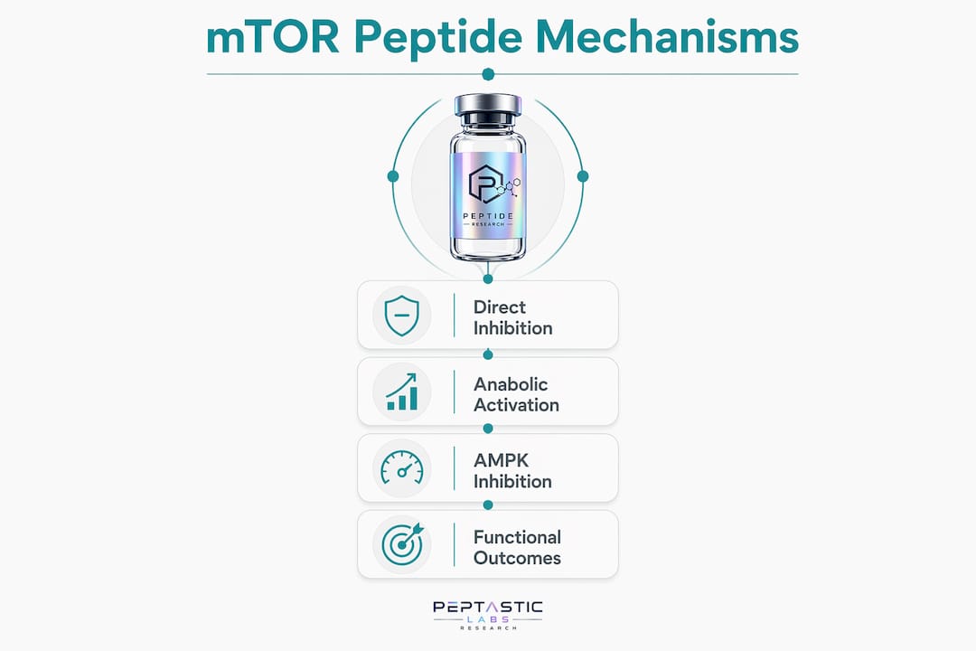
The 2026 literature has produced two findings that materially shift how researchers should interpret peptide-based mTOR modulation outcomes.
Inhibitory peptide design reaches near-native binding affinity
The mTOR-RHEB interface study published in april 2026 confirmed that mutational optimization of interfacial amino acid sequences produces peptides with binding energies within 1.3 kcal/mol of the native complex. This gap is small enough to suggest functional competitive inhibition in cellular assays. The finding validates computational peptide design as a primary, not supplementary, tool in mTORC1 inhibitor development.
MOTS-c reveals a metabolic-functional paradox
A june 2026 study on mesenchymal stem cells (MSCs) from obese donors (BMI ≥30 kg/m²; n=6 per group) showed that exogenous MOTS-c restored mitochondrial metabolic signaling but simultaneously reduced MSC proliferation and increased senescence markers. That result is counterintuitive. Metabolic pathway activation did not translate to improved reparative function. The data separates two outcomes that researchers often conflate: metabolic signal restoration and cellular functional recovery.
| Peptide | mTOR Effect | Metabolic Outcome | Functional Outcome |
|---|---|---|---|
| MOTS-c | AMPK activation, mTORC1 inhibition | Restored mitochondrial signaling | Reduced proliferation, increased senescence |
| Sermorelin | mTORC1 activation via GH-IGF-1 | Increased anabolic signaling | Protein synthesis, tissue repair |
| Interface peptides (mTOR-RHEB) | Direct mTORC1 disruption | Reduced proliferative signaling | Potential antitumor effect |
The MOTS-c paradox has direct implications for study design. Researchers using metabolic pathway activation as a proxy for cellular health will draw incorrect conclusions. Functional assays, including proliferation, senescence staining, and differentiation capacity, must accompany metabolic readouts in every mTOR peptide study.
- Metabolic signal restoration does not equal functional cellular improvement
- Senescence markers must be measured alongside AMPK and mTOR phosphorylation status
- Obesity context (BMI ≥30 kg/m²) modifies peptide outcomes, requiring stratified experimental designs
How does mTOR peptide research inform disease mechanisms and therapy?
mTORC1 hyperactivation contributes to cancer, type 2 diabetes, and aging-related pathologies. Peptide research provides a mechanistic lens for understanding how these diseases emerge from deregulated mTOR signaling, and a design framework for therapeutic intervention.
Cancer and proliferative disease
Uncontrolled mTORC1 activity drives tumor cell protein synthesis and suppresses autophagy-mediated quality control. Peptides that disrupt the mTOR-RHEB interface represent a structurally novel cancer therapeutic strategy, distinct from rapamycin analogs (rapalogs) that bind FKBP12 and only partially inhibit mTORC1. Interface peptides target the activation step directly. That mechanistic difference matters because analog-resistant tumors may remain sensitive to interface disruption.
Metabolic disease and obesity
In metabolic disease models, mTOR inhibition via AMPK-activating peptides shifts cells away from anabolic excess and toward mitochondrial efficiency. The MOTS-c data from obese MSC donors illustrates both the promise and the complexity of this approach. Metabolic correction is achievable, but functional tissue repair requires additional signaling inputs beyond AMPK activation alone.
Longevity and aging
mTOR inhibition extends lifespan and improves disease markers in preclinical models. Peptides such as Epithalon connect telomere biology to mTOR signaling in aging contexts. BPC-157 activates pathways that converge on mTOR and has been studied for tissue repair and cytoprotective effects. The longevity research field treats mTOR suppression as a core mechanism, with peptide-based approaches offering tissue-specific modulation that systemic rapalogs cannot provide.
Pro Tip: In cancer and aging studies, distinguish between mTORC1-specific and dual mTORC1/mTORC2 inhibition. Many peptides act upstream and affect both complexes. Confirm complex-specific phosphorylation markers (S6K1 for mTORC1, Akt Ser473 for mTORC2) in every experiment.
What techniques are used to design peptides targeting mTOR?
Peptide design for mTOR modulation follows a structured pipeline: computational modeling, mutational optimization, binding validation, and functional assay confirmation. Skipping any stage produces compounds with unpredictable in-cell behavior.
- Identify the target interface. Map the contact residues between mTOR and its binding partners (RHEB, Raptor, Rictor) using crystal structure data from the Protein Data Bank.
- Extract interfacial sequences. Derive candidate peptide sequences from the amino acids that contribute most to binding energy at the interface.
- Run protein-peptide docking. Use HADDOCK 2.4 to model peptide-protein binding geometry and score intermolecular contacts including hydrogen bonds and hydrophobic interactions.
- Calculate binding energies with FoldX5.0. Quantify the free energy of binding for wild-type and mutated peptide variants. Target energies within 2 kcal/mol of the native interaction.
- Mutational optimization. Substitute interfacial residues systematically to improve affinity, reduce off-target contacts, and increase proteolytic stability.
- In vitro validation. Confirm binding with surface plasmon resonance or isothermal titration calorimetry. Measure downstream mTOR pathway markers (phospho-S6K1, phospho-4EBP1) in cell-based assays.
- Functional outcome assessment. Measure proliferation, senescence, autophagy flux, and differentiation capacity. Do not stop at phosphorylation readouts.
The HADDOCK and FoldX pipeline has become the standard entry point for academic mTOR peptide programs. Researchers who bypass computational pre-screening synthesize far more peptide variants than necessary, increasing cost and time without improving hit rates. Computational triage is the rate-limiting step that determines experimental efficiency.
Key Takeaways
The mTOR pathway integrates four critical inputs, and peptide research reveals that metabolic signal activation does not automatically produce functional cellular improvement, a distinction that must drive study design.
| Point | Details |
|---|---|
| mTOR complexes have distinct roles | mTORC1 controls anabolic synthesis; mTORC2 governs Akt phosphorylation and cytoskeletal organization. |
| Peptides engage mTOR through three nodes | Direct interface disruption, GH-IGF-1 axis activation, and AMPK-mediated inhibition are the three primary mechanisms. |
| Metabolic and functional outcomes diverge | MOTS-c restores mitochondrial signaling but reduces MSC proliferation, proving metabolic readouts alone are insufficient. |
| Computational design is the entry point | HADDOCK and FoldX binding energy calculations must precede synthesis to reduce failed candidates. |
| Disease context shapes peptide selection | Cancer, metabolic disease, and aging each require different mTOR modulation strategies and distinct validation endpoints. |
The complexity researchers underestimate
The mTOR field has a clarity problem that peptide research is beginning to expose. For years, the dominant assumption was that mTOR inhibition equals benefit: suppress the pathway, reduce cancer proliferation, extend lifespan, improve metabolic health. The MOTS-c data from june 2026 breaks that assumption cleanly. Restoring mitochondrial metabolic signaling in obese MSCs made the cells metabolically "correct" by phosphorylation standards and functionally worse by every reparative measure.
That finding should change how researchers design experiments. Phosphorylation cascades are not outcomes. They are intermediate signals. The actual outcomes are proliferation, differentiation, senescence, and tissue function. Any peptide study that reports only pathway activation data without functional endpoints is incomplete, regardless of how clean the Western blots look.
The second underappreciated complexity is context specificity. The same peptide produces different results in lean versus obese tissue, in young versus aged cells, and in cancer versus normal epithelium. Peptide signaling research cannot be conducted in a single cell line and generalized. The MOTS-c obesity context finding is a direct warning against that practice.
The opportunity here is real. Peptides offer tissue-targeted mTOR modulation that small molecules and biologics cannot match in specificity. But realizing that opportunity requires integrated study designs that combine computational modeling, biochemical validation, and functional cellular assays in physiologically relevant models. Researchers who build that pipeline now will produce findings that hold up in translational settings. Those who rely on pathway activation readouts alone will generate data that does not replicate.
— Tintastic
Peptasticlabs: research-grade peptides for mTOR pathway studies
Researchers studying mTOR signaling need compounds with verified purity and complete batch documentation. Peptasticlabs supplies over 22 independently tested peptides, each verified to ≥99% purity via HPLC, with Certificates of Analysis available on request.

The catalog includes MOTS-c for AMPK and mitochondrial signaling studies, BPC-157 for mTOR-convergent tissue repair research, and GH secretagogues including Sermorelin for anabolic mTOR activation models. Every compound ships with third-party verification and full batch documentation. Researchers can browse the full catalog or visit Peptasticlabs to review compound specifications and request documentation before ordering.
FAQ
What is the mTOR pathway's primary function in cells?
mTOR integrates nutrient, energy, growth factor, and stress signals to coordinate protein synthesis, lipid production, and autophagy. mTORC1 drives anabolic processes; mTORC2 regulates Akt and cytoskeletal dynamics.
How does mTOR affect peptide-based research outcomes?
The mTOR pathway determines whether a peptide produces anabolic or catabolic cellular effects. Peptides targeting opposing mTOR nodes produce fundamentally different metabolic and functional outcomes, requiring researchers to define their signaling objective before compound selection.
What is the significance of the mTOR-RHEB interface in peptide design?
The mTOR-RHEB interface is the activation contact point for mTORC1. Peptides designed from this interface can competitively inhibit mTORC1 with binding energies near the native complex, offering a structurally distinct inhibition mechanism compared to rapalogs.
Can metabolic pathway activation predict functional cellular improvement?
No. The MOTS-c study in obese MSCs showed that AMPK activation and metabolic signal restoration did not improve reparative function and increased senescence markers. Functional assays are required alongside phosphorylation readouts.
Which peptide classes are most studied for mTOR modulation?
GH secretagogues, AMPK-activating mitochondrial peptides such as MOTS-c, interface-derived inhibitory peptides, and IGF-1 analogs represent the four primary classes. Each engages mTOR at a different node and requires distinct validation endpoints.