
In vitro peptide testing is defined as the systematic laboratory evaluation of peptide compounds using analytical and functional assays to determine identity, purity, concentration, and biological activity. The field relies on two distinct assay categories: analytical assays that answer "what is present and how much," and bioassays that answer "what does it do." Techniques including reversed-phase HPLC, LC-MS/MS, surface plasmon resonance (SPR), and ELISA form the core methodological toolkit for researchers characterizing peptides across metabolic, cognitive, tissue repair, and immunology applications. Peptasticlabs verifies each compound in its catalog to ≥99% purity via HPLC, reflecting the standards this article covers.
What is in vitro peptide testing and how do the two assay types differ?
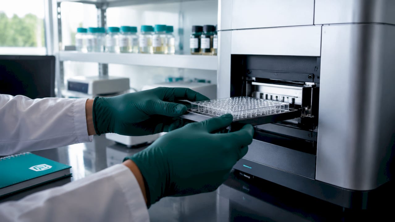
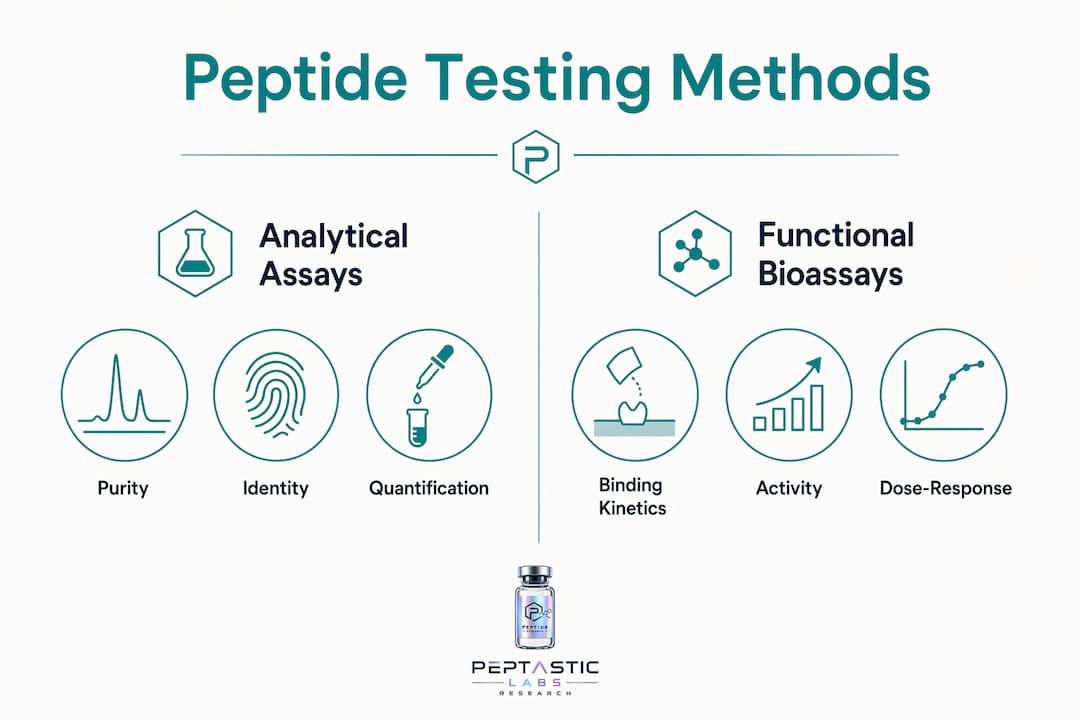
Analytical assays and bioassays serve fundamentally different purposes, and conflating them is one of the most common errors in peptide research design. Analytical assays characterize the physical and chemical properties of a peptide sample. Bioassays evaluate how that peptide interacts with biological systems. Both are required for complete characterization, and neither substitutes for the other.
Analytical assays confirm identity, measure purity, and quantify concentration. Bioassays assess receptor binding kinetics, cellular signaling, proliferation, migration, and cytotoxicity. A peptide can be analytically pure at ≥99% and still show no functional activity in a relevant cell model. Conversely, a peptide with trace impurities can produce confounding bioassay signals that obscure true activity.
Understanding assay purpose before designing a study determines which controls, concentrations, and readouts are appropriate. Researchers who skip this distinction often generate data that cannot be interpreted cleanly or reproduced across labs.
What analytical assays are used in in vitro peptide analysis?
Reversed-phase HPLC is the standard method for peptide purity determination and quantitation. The technique separates peptide species by hydrophobicity, and UV detection at 214–220 nm captures the peptide bond absorbance. Purity is reported as the percentage of the main peak area relative to total peak area. For quantitation, HPLC-UV is calibrated against a reference standard of known concentration.
LC-MS/MS extends HPLC by coupling mass spectrometric detection for identity confirmation. The technique confirms molecular weight and sequence fragments simultaneously. Multiple reaction monitoring (MRM) on triple-quadrupole instruments is the standard approach for quantitation in complex biological matrices such as plasma or cell lysate. Isotope-labeled internal standards are added to correct for matrix effects and instrument variability.
Key parameters measured in analytical in vitro peptide analysis include:
- Purity (%): Main peak area by HPLC-UV, target ≥99% for research-grade material
- Molecular weight: Confirmed by LC-MS/MS against theoretical mass
- Quantitation: Calibrated HPLC-UV or MRM with internal standards
- Counterion content: Assessed by ion chromatography or titration
- Residual solvents: Detected by headspace GC where required
Sample throughput is a practical constraint in analytical workflows. Multiplexing cyclic peptides in a single analytical injection using instruments such as the Thermo Scientific Orbitrap Astral reduces instrument time while preserving data quality. This strategy is particularly useful for stability and biotransformation studies where multiple time points must be analyzed.
| Parameter | Method | Key Output |
|---|---|---|
| Purity | Reversed-phase HPLC-UV | % main peak area |
| Identity | LC-MS/MS | Molecular weight, sequence fragments |
| Quantitation | MRM on triple-quadrupole | Concentration in matrix |
| Counterion | Ion chromatography | TFA or chloride content |
Pro Tip: Residual trifluoroacetic acid (TFA) from solid-phase synthesis is a frequent source of analytical error. TFA alters peptide conformation and membrane permeability, which distorts both purity readings and downstream bioassay results. Exchange TFA to physiological counterions such as chloride using validated protocols before running any functional assay.
What functional bioassays evaluate peptide activity in vitro?
Functional bioassays measure what a peptide does in a biological context. The three most widely used formats are SPR, ELISA, and cell culture-based potency assays. Each format answers a different question about peptide behavior.
SPR quantifies binding kinetics in real time. The peptide flows over an immobilized target protein, and mass increase on the sensor surface is detected as a shift in resonance angle. SPR outputs association rate (ka), dissociation rate (kd), and equilibrium dissociation constant (KD). These parameters define binding affinity and residence time, both of which are critical for understanding pharmacological potential.
ELISA formats use enzyme-conjugated detection antibodies, typically horseradish peroxidase (HRP) or alkaline phosphatase (AP), to quantify binding or competitive displacement. ELISA is lower throughput than SPR but accessible to most labs and well-suited for screening multiple peptide variants against a single target.
Cell culture-based potency assays measure functional responses in living cells. Common readouts include:
- cAMP accumulation: Measures GPCR agonist or antagonist activity
- Tube formation: Assesses pro-angiogenic peptides in endothelial cell models
- Wound migration: Quantifies cell motility responses to peptide treatment
- Cytotoxicity: Detects membrane disruption or apoptosis induction
- Proliferation: Measures mitogenic or anti-proliferative effects
Cell line selection is not arbitrary. Dose-response designs use cell-specific models matched to the peptide's target, such as pancreatic beta cells for metabolic peptides or neuronal cell lines for cognitive research compounds. Using an irrelevant cell line produces data that cannot be interpreted in the context of the intended biology.
Cell culture programs for peptide development follow a defined progression: hit confirmation, structure-activity relationship optimization, mechanism studies, and lead selection. Each stage uses the same cell model to maintain comparability across the program.
Pro Tip: Testing across 10–12 concentration points spanning 4–6 log units is the minimum required to derive reliable EC50 and IC50 values. Single-point bioassays are insufficient. They miss the full dose-response curve and can produce misleading potency estimates that do not replicate.
How do practical factors affect in vitro testing outcomes?
Sample preparation and formulation decisions made before the assay runs determine data quality as much as the assay itself. Three factors consistently affect reproducibility: counterion content, peptide stability in the assay matrix, and cell line suitability.
Residual TFA is the most underestimated variable in peptide bioassays. TFA alters conformation and membrane permeability, producing false signals in cell-based assays and mass measurement errors in LC-MS/MS. Validated exchange protocols use 10 mM HCl to remove TFA without affecting peptide purity. Labs that skip counterion exchange introduce a systematic error that cannot be corrected after the fact.
Practical steps for reliable in vitro peptide testing:
- Confirm purity by HPLC before any bioassay. Impure samples produce uninterpretable functional data.
- Exchange TFA to chloride using validated ion-exchange protocols before cell-based work.
- Prepare fresh stock solutions in appropriate solvents (DMSO or aqueous buffer) and verify concentration by UV or LC-MS/MS.
- Select the cell line based on target expression, not convenience.
- Include validated control compounds at each assay run to confirm system performance.
- Run dose-response designs rather than single-point tests for all potency measurements.
Multiplexing strategies also improve throughput without sacrificing quality. Pooling peptide samples for combined stability and biotransformation analysis in a single MS injection reduces instrument time and increases the number of compounds characterized per run. This approach is standard in high-throughput drug discovery workflows and applicable to academic research programs with limited instrument access.
Pro Tip: Certificates of Analysis (CoA) from your peptide supplier should document HPLC purity, LC-MS/MS identity confirmation, and counterion content. A CoA for peptide research that lacks these three data points is insufficient for assay design decisions.
What applications and limitations should researchers know?
In vitro peptide testing supports research across drug discovery, mechanistic biology, and formulation development. The range of peptide applications spans metabolic signaling, immune modulation, tissue repair, and cognitive pathway studies. Each application requires a matched assay format: receptor binding studies use SPR or competitive ELISA, while cellular mechanism studies require relevant cell-based potency assays.
The primary limitation of in vitro data is its inability to predict clinical outcomes. In vitro results from animal and cell models do not guarantee human clinical benefit. Peptide stability, bioavailability, and target access in a living organism introduce variables that no cell culture model captures. This gap is not a failure of in vitro methods. It is a structural limitation that requires in vitro data to be treated as hypothesis-generating rather than conclusive.
Researchers should apply the following practices to maintain data integrity:
- Use validated control compounds at every assay run to confirm assay performance and flag drift
- Replicate across independent experiments, not just technical replicates within a single run
- Document all assay conditions including passage number, serum lot, and incubation time
- Interpret EC50 and IC50 values in the context of the assay format, not as absolute potency claims
- Apply statistical rigor with appropriate sample sizes and repeated assay designs
Control compounds in peptide studies serve as the internal benchmark that separates genuine peptide activity from assay noise. Omitting them is the single fastest way to generate irreproducible data.
Key Takeaways
In vitro peptide testing requires both analytical assays for chemical characterization and functional bioassays for biological activity, and neither category alone constitutes complete peptide profiling.
| Point | Details |
|---|---|
| Two assay categories required | Analytical assays confirm identity and purity; bioassays measure functional activity. |
| TFA removal is non-negotiable | Residual TFA distorts both mass measurements and cell-based assay results. |
| Dose-response over single-point | Testing 10–12 concentration points across 4–6 log units is required for valid EC50 and IC50 values. |
| Cell line selection determines relevance | Assay cells must express the peptide's target to produce interpretable data. |
| In vitro data is hypothesis-generating | Cell and animal model results do not predict human clinical outcomes. |
Why I think most peptide assay failures happen before the instrument is turned on
Most reproducibility problems in peptide research trace back to decisions made at the bench before any instrument runs. Researchers focus on assay optimization, software settings, and statistical analysis, but the real failure points are upstream: peptide purity not confirmed, TFA not exchanged, stock concentration not verified, cell line not validated for target expression.
The field has excellent analytical tools. HPLC, LC-MS/MS, SPR, and cell-based potency assays are well-characterized and widely available. What is less well-distributed is the discipline to use them in sequence. Analytical data should gate entry to bioassays. A peptide that has not been confirmed at ≥99% purity by HPLC has no business in a cell-based potency assay. The data it generates will not replicate, and the time spent running it is wasted.
Emerging multiplexing approaches using high-resolution mass spectrometry are genuinely useful for throughput. But they amplify good practice, not bad. Pooling impure or poorly characterized peptides for multiplex analysis produces high-throughput noise, not high-throughput insight.
The most underutilized resource in peptide research is the supplier's documentation. A full CoA with HPLC chromatogram, LC-MS/MS spectrum, and counterion data tells you whether your starting material is fit for purpose before you design a single experiment. Researchers who treat CoA review as a formality rather than a scientific step are setting themselves up for failed replication.
— Tintastic
Peptasticlabs supports your in vitro peptide testing workflow
Peptasticlabs supplies research-grade peptides verified to ≥99% purity via HPLC, with LC-MS/MS identity confirmation and full batch documentation available on request. Every compound in the catalog is independently tested before release, giving researchers a documented starting point for both analytical and functional assay work.

The peptide catalog covers metabolic, cognitive, tissue repair, longevity, immunology, and cosmetic research applications, with Certificates of Analysis available for each batch. Researchers who need verified, documented compounds for cell-based potency assays, SPR binding studies, or LC-MS/MS quantitation work will find the full product range and supporting documentation at Peptasticlabs.
FAQ
What is in vitro peptide testing?
In vitro peptide testing is the laboratory-based evaluation of peptide compounds using analytical assays (HPLC, LC-MS/MS) to confirm identity and purity, and functional bioassays (SPR, ELISA, cell-based assays) to measure biological activity.
What are peptides used for in research?
Peptides are used to study metabolic signaling, immune modulation, tissue repair, cognitive pathways, and receptor pharmacology across drug discovery and mechanistic biology programs.
How is peptide purity measured in vitro?
Purity is measured by reversed-phase HPLC-UV, reported as the main peak area percentage. LC-MS/MS confirms molecular identity alongside purity data.
Why does TFA affect in vitro peptide assay results?
Residual TFA from synthesis alters peptide conformation and membrane permeability, producing false signals in cell-based assays and mass measurement errors in LC-MS/MS. Ion exchange to chloride before testing removes this interference.
Can in vitro peptide results predict human clinical outcomes?
In vitro results from cell and animal models do not guarantee human clinical benefit. These results are hypothesis-generating and require further validation before clinical conclusions can be drawn.