
IGF-1 coordinates nutrient-driven anabolic and survival programs primarily through two canonical routes: PI3K→Akt→mTOR, which governs protein synthesis, glucose uptake, and anti-apoptotic signaling, and Ras→MAPK (ERK), which drives proliferative and differentiation programs. Both cascades initiate when IGF-1 binds IGF-1R or IGF-1R/IR hybrid receptors, triggering autophosphorylation of the receptor's beta subunits and recruitment of IRS-1/2 and Shc adaptors. The schematic is direct: IGF-1 → IGF-1R/hybrid receptors → autophosphorylation → IRS/Shc → (1) PI3K→Akt→mTOR → metabolic readouts; (2) Ras→MAPK → growth/differentiation.
Researchers designing metabolic experiments should expect these primary readouts from IGF-1 signaling pathways:
- Glucose uptake: GLUT4 translocation in muscle and adipose tissue
- Hepatic gluconeogenesis suppression: FOXO1 phosphorylation reducing G6Pase and PEPCK expression
- Fatty acid handling: altered FA uptake and oxidation, particularly in skeletal muscle via CD36
- Protein synthesis: mTORC1-driven S6K and 4E-BP1 phosphorylation
- Anti-apoptosis: Akt-mediated BAD phosphorylation and FOXO nuclear exclusion
Table of Contents
- How is IGF-1 produced, circulated, and regulated?
- What is the structure and activation mechanism of IGF-1R?
- How do PI3K→Akt→mTOR and Ras→MAPK transduce IGF-1 signals?
- How does IGF-1 cross-talk with insulin signaling drive metabolic disease?
- How does IGF-1 act differently across liver, muscle, adipose tissue, pancreas, and hypothalamus?
- How do IGFBPs regulate IGF-1 bioavailability?
- What do genetic mouse models reveal about IGF-1 metabolic function?
- What are the translational implications for metabolic disease and cancer?
- How do you disentangle IGF-1 versus insulin effects experimentally?
- What should researchers remember about IGF-1 metabolic signaling?
- Key Takeaways
- The open questions that matter most in IGF-1 metabolic research
- Research-grade IGF-1 reagents with full documentation
- Useful sources
How is IGF-1 produced, circulated, and regulated?
The liver is the dominant source of circulating IGF-1, synthesizing and secreting it under direct growth hormone (GH) stimulation. GH binds hepatic GH receptors, activates JAK2/STAT5 signaling, and drives IGF-1 gene transcription. Paracrine and autocrine synthesis occurs in skeletal muscle, bone, and other tissues, where locally produced IGF-1 acts independently of circulating levels.
Most circulating IGF-1 does not travel free. Hepatic IGF-1 production results in peptide that is predominantly bound to IGFBPs, particularly IGFBP-3, and the acid-labile subunit (ALS), forming ternary complexes that extend the half-life from minutes to hours and restrict tissue access. Only a small free fraction is immediately bioavailable to receptors.
Several systemic variables modulate circulating IGF-1 levels:
- GH pulse amplitude and frequency: the primary driver of hepatic IGF-1 output; GH deficiency sharply reduces circulating IGF-1
- Nutritional status: caloric restriction and protein deficiency suppress hepatic IGF-1 despite normal GH secretion, uncoupling the GH/IGF-1 axis
- Age: IGF-1 peaks in puberty and declines progressively through adulthood
- Insulin: required for hepatic GH receptor expression; portal insulin deficiency (as in T1DM) reduces IGF-1 even when GH is elevated
- Inflammation and chronic disease: cytokines (IL-6, TNF-α) suppress hepatic IGF-1 synthesis; obesity and T2DM alter the axis through insulin resistance and hyperinsulinemia.
Assay note: Sampling conditions alter measured IGF-1 substantially. Fasting reduces free IGF-1 by increasing IGFBP-1, while GH pulses transiently raise total IGF-1. Assays measuring total IGF-1 after acid-ethanol extraction report different values than those measuring free peptide directly. Document feeding state, time of day, and assay format in every experiment.
What is the structure and activation mechanism of IGF-1R?
IGF-1R is a receptor tyrosine kinase (RTK) assembled as a disulfide-linked (αβ)₂ heterotetramer. The two extracellular alpha subunits contain the ligand-binding domains; the two transmembrane beta subunits carry the intracellular tyrosine kinase domains. Ligand binding to the alpha subunit induces a conformational change in the beta subunit that enables trans-autophosphorylation at key tyrosine residues in the activation loop (Y1135/Y1136 in human IGF-1R), fully activating kinase activity.

Adaptor recruitment and pathway bifurcation
Activated IGF-1R phosphorylates two principal adaptor classes:
- IRS-1 and IRS-2: docking proteins with multiple tyrosine phosphorylation sites that recruit the p85 regulatory subunit of PI3K, directing signaling toward the PI3K→Akt→mTOR metabolic axis
- Shc: an adaptor that recruits Grb2/SOS, activating Ras and directing signaling toward the Raf→MEK→ERK proliferative axis
The relative engagement of IRS versus Shc determines whether a given cell responds with a predominantly metabolic or mitogenic program, and this balance shifts with receptor expression level, ligand concentration, and cellular context.
Hybrid receptor biology and experimental consequences
In tissues co-expressing IGF-1R and insulin receptor (IR), hemireceptors can assemble into IGF-1R/IR hybrids. Hybrid receptors bind IGF-1 with higher affinity than insulin, meaning that some effects attributed to IGF-1R homodimers in vivo may actually be mediated through hybrids. This has direct experimental consequences:
- Receptor-specific antibodies and inhibitors may not fully block hybrid-mediated signaling
- Cell lines with high IR expression (e.g., hepatocytes, adipocytes) will have substantial hybrid populations
- Knockdown of IGF-1R reduces hybrid abundance and may alter insulin sensitivity independently of IGF-1 signaling
- Cryo-EM structural data have clarified how engineered ligands can bias activation toward IGF-1R versus IR, offering a strategy for cleaner mechanistic experiments
Researchers should characterize hybrid receptor abundance in their model system before attributing metabolic effects exclusively to IGF-1R.
How do PI3K→Akt→mTOR and Ras→MAPK transduce IGF-1 signals?
The two canonical cascades diverge at the IRS/Shc branch point and converge on distinct but overlapping metabolic programs.

PI3K→Akt→mTOR: the metabolic axis
IRS-1/2 phosphorylation recruits the p85/p110 PI3K heterodimer, which phosphorylates PIP2 to generate PIP3 at the plasma membrane. PIP3 recruits PDK1 and Akt (PKB), enabling PDK1 to phosphorylate Akt at T308 and mTORC2 to phosphorylate Akt at S473, achieving full activation. Akt then phosphorylates multiple substrates with direct metabolic consequences:
- TSC2 inhibition: releases mTORC1 from inhibition, activating S6K1 and 4E-BP1 to drive cap-dependent translation and protein synthesis
- FOXO1/3 phosphorylation: excludes FOXO transcription factors from the nucleus, suppressing G6Pase and PEPCK transcription and reducing hepatic gluconeogenesis
- AS160 phosphorylation: releases the Rab-GAP brake on GLUT4 vesicle trafficking, enabling glucose uptake in muscle and adipose tissue
- GSK-3β inhibition: promotes glycogen synthesis
The mTOR pathway also integrates nutrient sensing: amino acid sufficiency activates Rag GTPases that recruit mTORC1 to the lysosomal surface, where it is activated by Rheb. IGF-1 signaling through Akt converges on this same mTORC1 node, making the pathway a hub for both hormonal and nutritional inputs.
Ras→MAPK (ERK): the proliferative axis
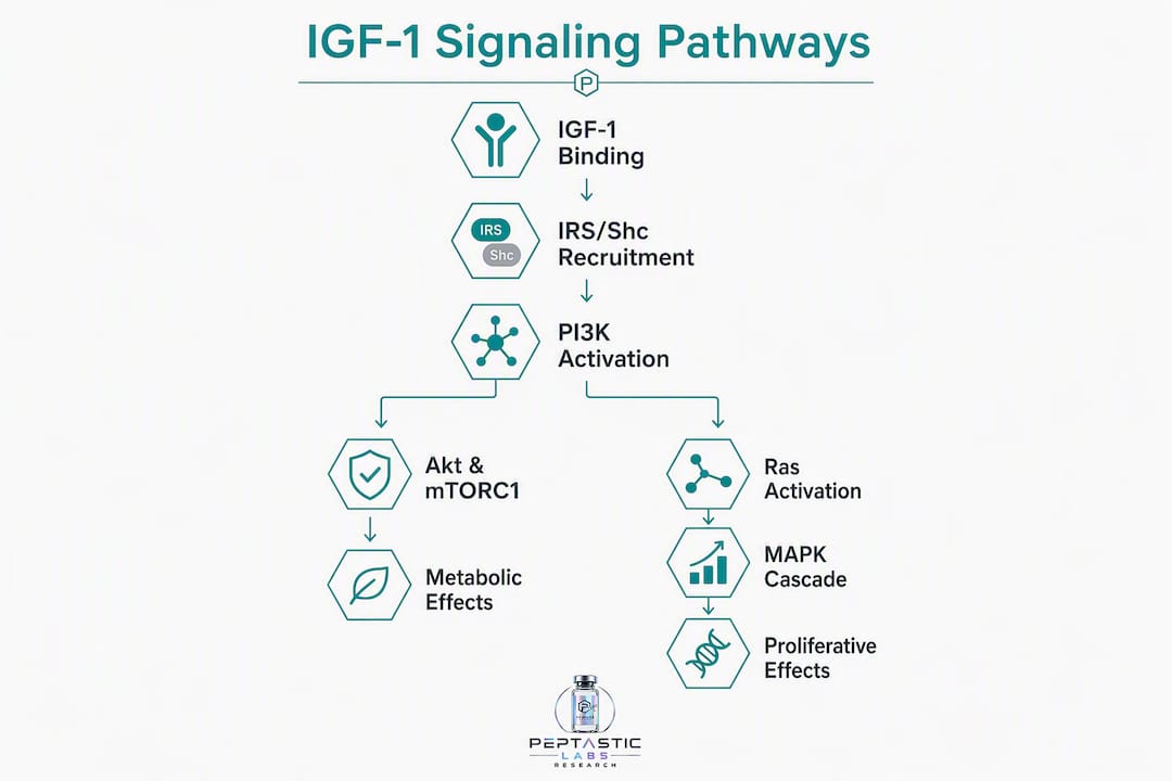
Shc phosphorylation recruits Grb2, which binds the guanine nucleotide exchange factor SOS, activating Ras. GTP-loaded Ras activates Raf, which phosphorylates MEK1/2, which in turn phosphorylates ERK1/2. Nuclear ERK drives transcription of immediate-early genes (c-Fos, c-Jun, Egr-1) and activates RSK, linking IGF-1 stimulation to cell cycle entry and growth-related metabolic reprogramming.
Pathway nodes, metabolic readouts, and standard assays
| Pathway Node | Metabolic Readout | Standard Assay |
|---|---|---|
| pAkt (S473 / T308) | Glucose uptake, protein synthesis, anti-apoptosis | Phospho-western blot, ELISA |
| pFOXO1 | Hepatic gluconeogenesis suppression | ChIP, G6Pase/PEPCK mRNA qPCR |
| pS6K1 / p4E-BP1 | Cap-dependent translation, protein synthesis | Phospho-western blot |
| pGSK-3β | Glycogen synthesis | Glycogen assay, phospho-blot |
| pAS160 | GLUT4 translocation, glucose uptake | GLUT4 translocation assay, 2-DG uptake |
| pERK1/2 | Proliferation, transcriptional reprogramming | Phospho-western blot, reporter assay |
Pro Tip: When running phospho-blots for IGF-1 signaling, always probe for both Akt S473 and T308 simultaneously. S473 alone can be elevated by mTORC2 independently of PI3K input; T308 phosphorylation confirms PDK1-dependent, PI3K-driven Akt activation.
How does IGF-1 cross-talk with insulin signaling drive metabolic disease?
IGF-1 and insulin share the most structurally conserved receptor-ligand pair in the RTK superfamily. Both receptors signal through IRS-1/2 and PI3K/Akt, and at high concentrations each ligand can activate the other's receptor. This overlap is not merely academic: it shapes how the IGF axis behaves in metabolic disease.
Key mechanistic overlaps and divergences:
- Shared nodes: IRS-1/2, PI3K p85/p110, Akt, mTORC1, FOXO1, GSK-3β
- Divergence points: tissue receptor expression (IGF-1R predominates in muscle and brain; IR predominates in liver and adipose tissue in adults), receptor affinity for each ligand, and downstream transcriptional targets
- Hybrid receptors: in tissues co-expressing both receptors, hybrids bind IGF-1 preferentially, meaning IGF-1 can activate IR-associated signaling nodes even when IR homodimer occupancy is low
The most clinically relevant cross-talk mechanism is S6K-mediated negative feedback. Chronic mTORC1/S6K1 activation, whether from IGF-1, insulin, or nutrient excess, drives serine phosphorylation of IRS-1 at inhibitory sites (S307, S636/639 in rodents). This reduces IRS-1 tyrosine phosphorylation in response to subsequent insulin or IGF-1 stimulation, attenuating PI3K recruitment and creating a state of receptor-level resistance. In obesity and T2DM, chronic hyperinsulinemia and nutrient excess sustain this feedback loop, progressively degrading both insulin and IGF-1 signaling efficiency.
The metabolic consequences extend beyond glucose handling. In obesity and T2DM, the coordinated IGF-1/insulin response fails to suppress hepatic gluconeogenesis or drive peripheral glucose uptake effectively. Elevated free fatty acids further impair IRS signaling through PKC-θ-mediated serine phosphorylation, compounding the defect. The result is a state where both hormones are present but their shared downstream pathway is blunted.
Cancer risk represents an additional dimension of this cross-talk. Hyperinsulinemia reduces IGFBP-1 and IGFBP-2 production, increasing free IGF-1. Elevated free IGF-1, acting through IGF-1R and hybrids, sustains Akt and ERK activity in proliferating cells, linking the metabolic syndrome phenotype to increased mitogenic drive.
How does IGF-1 act differently across liver, muscle, adipose tissue, pancreas, and hypothalamus?
Liver
The liver is both the primary source and a major target of IGF-1 signaling. Hepatic IGF-1 acts in an autocrine/paracrine manner to suppress gluconeogenesis via FOXO1 phosphorylation, reducing G6Pase and PEPCK expression. Liver-specific IGF-1 knockout (LID) mice show elevated GH and reduced circulating IGF-1, with compensatory hyperinsulinemia and mild insulin resistance, confirming the liver's role in maintaining systemic IGF-1 tone. Standard endpoints: G6Pase and PEPCK mRNA by qPCR, hyperinsulinemic-euglycemic clamp, and hepatic glucose output by tracer dilution.
Skeletal muscle
Skeletal muscle is the primary site of IGF-1-driven protein anabolism. IGF-1 activates PI3K/Akt/mTORC1 to phosphorylate S6K1 and 4E-BP1, stimulating protein synthesis and inhibiting atrophy pathways including atrogin-1 and MuRF1. Muscle also responds to IGF-1 with increased CD36-mediated fatty acid uptake and oxidation, contributing to systemic lipid clearance. Because skeletal muscle accounts for the majority of insulin-stimulated glucose disposal, IGF-1's effects on muscle IGF-1R and hybrid receptors have outsized implications for whole-body insulin sensitivity. Relevant assays include metabolic research peptide protocols for protein synthesis (puromycin incorporation, ³H-phenylalanine), GLUT4 translocation, and ex vivo glucose uptake.
Adipose tissue
Adipose IGF-1 signaling shifts substantially with differentiation state. Preadipocytes express high IGF-1R and respond to IGF-1 with differentiation signals; as cells mature into adipocytes, IGF-1R expression declines and IR dominance increases. At physiologic IGF-1 concentrations, mature adipocytes respond minimally to IGF-1 for lipid metabolism; supraphysiologic concentrations are required to drive lipolysis or lipogenesis directly. Brown and white adipose tissue differ further: brown adipocytes retain higher IGF-1R expression and respond to IGF-1 with thermogenic gene programs, relevant to the FIGIRKO phenotype discussed in the genetic models section. For tissue-specific adipose research, the role of peptides in adipose tissue provides useful experimental context.
Pancreas
In pancreatic beta cells, IGF-1 acts in a paracrine/autocrine manner to support beta-cell mass and function. IGF-1R activation promotes beta-cell survival via Akt-mediated BAD phosphorylation and FOXO1 exclusion, and supports glucose-stimulated insulin secretion by maintaining mitochondrial function. Beta-cell-specific IGF-1R knockout models show reduced beta-cell mass and impaired first-phase insulin secretion, confirming a direct trophic role. Relevant endpoints: beta-cell mass morphometry, GSIS assay, and Akt/FOXO1 phosphorylation in isolated islets.
Hypothalamus
Hypothalamic IGF-1 signaling participates in nutrient sensing and energy homeostasis. IGF-1R is expressed in arcuate nucleus neurons, where IGF-1 modulates neuropeptide Y (NPY) and POMC expression, influencing food intake and energy expenditure. Central IGF-1 also interacts with leptin signaling pathways, and hypothalamic IGF-1 resistance has been proposed as a contributor to the hyperphagia and reduced energy expenditure seen in diet-induced obesity. Behavioral endpoints include food intake monitoring, indirect calorimetry, and hypothalamic phospho-Akt immunohistochemistry.
How do IGFBPs regulate IGF-1 bioavailability?
Six IGF binding proteins (IGFBP-1 through IGFBP-6) modulate IGF-1 availability with distinct tissue distributions and regulatory functions. Their collective effect is to buffer free IGF-1 concentration, preventing receptor saturation under normal conditions while enabling rapid local release in response to proteolytic or hormonal signals.
- IGFBP-3: the dominant circulating carrier; forms ternary complexes with IGF-1 and ALS, extending IGF-1 half-life from minutes to roughly 12–16 hours and restricting vascular egress
- IGFBP-1: acutely regulated by insulin (suppressed) and glucagon (induced); rises rapidly with fasting, reducing free IGF-1; a sensitive marker of portal insulin action
- IGFBP-2: expressed in liver and CNS; inversely correlated with insulin sensitivity; elevated in T2DM and obesity
- IGFBP-4: inhibitory; cleaved by PAPP-A protease, releasing free IGF-1 locally in bone and vascular tissue
- IGFBP-5: can potentiate IGF-1 action in bone; also has IGF-independent effects
- IGFBP-6: preferentially binds IGF-2; relevant in CNS and tumor biology
Proteolytic cleavage of IGFBPs, particularly IGFBP-3 by pregnancy-associated plasma protein-A (PAPP-A) and IGFBP-4 by the same protease, releases free IGF-1 at the tissue level, creating a local bioavailability gradient independent of circulating total IGF-1. This mechanism is particularly active in wound healing, vascular remodeling, and follicular development.
Experimental caution: Total IGF-1 ELISAs after acid-ethanol extraction measure the sum of bound and free peptide and can mask acute changes in bioavailability. Fasting, acute illness, or insulin manipulation alters the free fraction substantially without changing total IGF-1. For metabolic studies, measure IGFBP-1 and IGFBP-3 alongside total IGF-1, and consider free IGF-1 assays (ultrafiltration-based or two-site immunoassay) when the research question involves acute metabolic perturbations. Document feeding state and sampling time in every experiment.
What do genetic mouse models reveal about IGF-1 metabolic function?
Transgenic and tissue-specific knockout models have been the primary tools for dissecting IGF-1's metabolic roles in vivo. The table below catalogs the most informative models, their phenotypes, and the metabolic insights they provide.
| Model | Genotype | Key Phenotype | Metabolic Insight |
|---|---|---|---|
| Global IGF-1 KO | Igf1⁻/⁻ | Severe growth retardation, perinatal lethality in many lines | Confirms IGF-1 as essential for postnatal growth; metabolic phenotype confounded by developmental defects |
| LID (liver IGF-1 KO) | Alb-Cre × Igf1^fl/fl | large reduction in circulating IGF-1; elevated GH; compensatory hyperinsulinemia | Liver-derived IGF-1 maintains systemic GH suppression and contributes to insulin sensitivity |
| Muscle IGF-1R KO | MCK-Cre × Igf1r^fl/fl | Reduced muscle mass; impaired glucose uptake in muscle | IGF-1R in muscle required for full anabolic response and glucose disposal |
| FIRKO (fat IR KO) | aP2-Cre × Insr^fl/fl | Protected from obesity-related glucose intolerance; increased lifespan | Adipose IR signaling paradoxically promotes metabolic dysfunction; its absence shifts energy balance favorably |
| FIGIRKO (fat IGF-1R + IR DKO) | aP2-Cre × Igf1r^fl/fl × Insr^fl/fl | Thermogenesis defects; altered energy expenditure; lipodystrophy | Both receptors required for normal adipose thermogenic function; IGF-1R contributes to brown fat programming |
| Beta-cell IGF-1R KO | RIP-Cre × Igf1r^fl/fl | Reduced beta-cell mass; impaired GSIS | IGF-1R supports beta-cell survival and secretory function |
| PAPP-A KO | Pappaⁿ/⁻ | Reduced local free IGF-1; extended lifespan in some studies | IGFBP proteolysis controls tissue-level IGF-1 bioavailability independently of circulating levels |
Tissue-specific knockout phenotypes such as FIRKO and FIGIRKO illustrate that metabolic outcomes depend on which receptor is absent in which tissue, not simply on total IGF-1 or insulin signaling levels. The FIRKO protection phenotype is counterintuitive: removing insulin receptor from fat improves systemic glucose tolerance, suggesting that adipose IR-driven lipid storage and inflammatory signaling contribute to whole-body insulin resistance.
Methodological caveats
Developmental compensation is a persistent problem in constitutive knockouts. Global IGF-1 KO mice activate compensatory growth programs that obscure adult metabolic phenotypes. Inducible Cre systems (e.g., tamoxifen-inducible CreERT2) allow receptor deletion in adult animals, avoiding developmental artifacts. Additional caveats:
- Hybrid receptor loss: knocking out IGF-1R in a tissue also eliminates hybrid receptors, which may independently mediate insulin signaling; phenotypes attributed to IGF-1R loss may partly reflect hybrid receptor loss
- Off-target Cre activity: aP2-Cre and RIP-Cre lines have documented expression in non-target tissues; validate with reporter lines
- Diet and age matching: metabolic phenotypes in IGF-1 axis models are highly diet- and age-dependent; high-fat diet challenges and longitudinal metabolic phenotyping are required for complete interpretation
- Rescue constructs: re-expression of IGF-1R in the knocked-out tissue confirms phenotype specificity
What are the translational implications for metabolic disease and cancer?
The IGF axis sits at the intersection of metabolic disease and cancer biology, making it both a therapeutic target and a source of on-target metabolic risk when inhibited.
Therapeutic modalities
- IGF-1R monoclonal antibodies (e.g., figitumumab, cixutumumab, ganitumab): block ligand binding; tested in oncology trials with limited single-agent efficacy; cause hyperglycemia and elevated GH/IGF-1 due to feedback
- IGF-1R tyrosine kinase inhibitors: small-molecule ATP-competitive inhibitors; cross-react with IR at higher concentrations, producing insulin resistance and hyperglycemia as on-target metabolic side effects
- Dual IR/IGF-1R inhibitors: broader metabolic disruption; useful experimentally but carry significant clinical metabolic toxicity
- mTOR inhibitors (rapamycin, everolimus): downstream of both IGF-1R and IR; suppress S6K-mediated feedback, paradoxically increasing IRS-1 signaling and Akt activity in some contexts; associated with hyperglycemia and dyslipidemia in clinical use
- Ligand sequestration: anti-IGF-1/IGF-2 antibodies or soluble IGF-1R decoys; reduce free ligand without direct receptor blockade
Clinical tradeoffs
IGF-1R inhibition in oncology trials has consistently produced hyperglycemia, elevated insulin, and in some cases frank insulin resistance, confirming the metabolic safety risk of disrupting this axis. The metabolic effects of GH and IGF-1 diverge importantly: GH is insulin-antagonizing, while IGF-1 is insulin-sensitizing. Blocking IGF-1R removes insulin-sensitizing tone, shifting the metabolic balance toward GH-driven insulin resistance.
For translational researchers, the practical implications are:
- Include glucose clamps, fasting glucose/insulin, and HbA1c as mandatory safety endpoints in any IGF-1R intervention study
- Monitor GH and free IGF-1 levels, not just total IGF-1, when assessing feedback responses to IGF-1R blockade
- Distinguish between IGF-1R homodimer blockade and hybrid receptor blockade when interpreting metabolic side effects
- Consider combination strategies that pair IGF-1R inhibition with insulin sensitizers (metformin, GLP-1 agonists) to manage metabolic toxicity
The cancer-metabolism intersection also runs in the other direction: hyperinsulinemia in metabolic syndrome reduces IGFBP-1/2, elevating free IGF-1 and sustaining mitogenic signaling in pre-malignant cells. Addressing insulin resistance may therefore reduce IGF-1-driven cancer risk without requiring direct IGF-1R blockade.
How do you disentangle IGF-1 versus insulin effects experimentally?
Separating direct IGF-1R-mediated effects from indirect insulin-mediated effects is one of the most common sources of interpretive error in IGF-1 metabolic research. The problem is structural: IGF-1 administration suppresses endogenous insulin secretion, meaning that many downstream metabolic readouts attributed to IGF-1 may be consequences of reduced insulin rather than direct IGF-1R activation.
A stepwise experimental checklist:
- Dose selection: use IGF-1 concentrations in the physiologic range (1–10 nM for in vitro) to avoid off-target IR activation; confirm receptor selectivity with a phospho-receptor antibody panel at each dose
- Paired insulin measurements: in every in vivo IGF-1 experiment, measure plasma insulin at the same timepoints as IGF-1 readouts; attribute effects to IGF-1R only when insulin is unchanged or controlled
- Fasting and feeding controls: standardize nutritional state before sampling; fasting alters IGFBP-1 and free IGF-1 independently of total IGF-1
- Receptor-selective pharmacology: use IGF-1R-selective inhibitors (e.g., NVP-AEW541 at low concentrations) rather than dual IR/IGF-1R inhibitors when the goal is to isolate IGF-1R contribution; confirm selectivity in your cell line
- Hybrid receptor characterization: immunoprecipitate with anti-IGF-1R and anti-IR antibodies and blot for the co-precipitated partner to quantify hybrid abundance in your model
- Tissue-specific knockout controls: where feasible, use inducible, tissue-restricted Cre models rather than global knockouts to avoid developmental compensation
- Tracer studies for hepatic gluconeogenesis: use ²H₂O or [U-¹³C]glucose tracer dilution to measure hepatic glucose output directly rather than inferring it from plasma glucose alone
- Lipid flux assays: ¹⁴C-palmitate oxidation and esterification assays in isolated muscle or adipose tissue provide direct readouts of FA handling independent of systemic hormonal changes
Recommended assay panel
| Assay | Target | Specificity Note |
|---|---|---|
| Phospho-Akt (S473 + T308) western | PI3K/Akt activation | T308 confirms PDK1/PI3K input; S473 alone insufficient |
| Phospho-ERK1/2 | MAPK activation | Confirm with MEK inhibitor control |
| Phospho-S6K1 | mTORC1 activity | Use rapamycin control to confirm mTOR dependence |
| Hybrid receptor IP | IGF-1R/IR hybrid abundance | Anti-IGF-1R IP + anti-IR blot |
| 2-DG glucose uptake | GLUT4-mediated uptake | Cytochalasin B control for non-transporter uptake |
| PEPCK / G6Pase qPCR | Gluconeogenic gene expression | Pair with FOXO1 ChIP |
| ¹⁴C-palmitate oxidation | FA oxidation | Tissue-specific; normalize to protein content |
Pro Tip: Reagent purity directly affects phospho-signaling reproducibility. Peptide preparations with sub-99% purity introduce contaminants that can activate stress kinases (JNK, p38) independently of IGF-1R, generating artifactual phospho-Akt or phospho-ERK signals. Always verify purity by HPLC and request a Certificate of Analysis before committing a peptide lot to a signaling experiment.
What should researchers remember about IGF-1 metabolic signaling?
The core claim: IGF-1 is a nutrient-state integrator that coordinates anabolic, anti-apoptotic, and glucose-regulatory programs through PI3K/Akt/mTOR and Ras/MAPK, with tissue-specific outcomes shaped by receptor expression, IGFBP-mediated bioavailability, and cross-talk with insulin signaling.
Key mechanistic anchors:
- PI3K/Akt/mTOR is the primary metabolic axis: glucose uptake, protein synthesis, gluconeogenesis suppression, and lipid handling all trace to Akt substrate phosphorylation
- Ras/MAPK drives proliferative and transcriptional programs; its metabolic contributions are secondary but relevant in cancer-metabolism contexts
- FOXO transcription factors are the principal transcriptional effectors of Akt in metabolic tissues; their phosphorylation state is a reliable readout of IGF-1/insulin axis activity
- S6K-mediated IRS serine phosphorylation creates a negative feedback loop that links chronic IGF-1/mTOR activation to insulin resistance
- Hybrid receptors are present in most metabolically relevant tissues and bind IGF-1 preferentially; effects attributed to IGF-1R homodimers may be partially or wholly hybrid-mediated
Common misinterpretations to avoid:
- Attributing all PI3K/Akt activation to IGF-1R when IR and hybrid receptors are co-expressed
- Interpreting reduced gluconeogenesis after IGF-1 administration as a direct IGF-1R effect without controlling for IGF-1-induced insulin suppression
- Using total IGF-1 ELISA as a proxy for bioavailable IGF-1 in acute metabolic studies
Rigorous experimental controls are not optional when assigning metabolic effects to IGF-1. Paired insulin measurements, receptor-selective pharmacology, and free IGF-1 quantification are the minimum standard for attributable mechanistic claims.
Key Takeaways
IGF-1 drives metabolic anabolism and glucose regulation through PI3K/Akt/mTOR and Ras/MAPK, with tissue-specific outcomes determined by receptor expression, IGFBP-mediated bioavailability, and cross-talk with insulin signaling.
| Point | Details |
|---|---|
| Dual canonical pathways | PI3K/Akt/mTOR governs metabolic readouts; Ras/MAPK drives proliferative programs; both initiate at IGF-1R autophosphorylation. |
| Hybrid receptors complicate attribution | IGF-1R/IR hybrids bind IGF-1 preferentially; characterize hybrid abundance before assigning effects to IGF-1R homodimers. |
| Free IGF-1 is the bioactive fraction | Total IGF-1 ELISA misses acute changes in bioavailability; measure IGFBP-1 and free IGF-1 alongside total in metabolic studies. |
| S6K feedback drives insulin resistance | Chronic mTORC1/S6K1 activation phosphorylates IRS-1 at inhibitory serine sites, degrading both IGF-1 and insulin signaling efficiency. |
| Peptasticlabs reagent standard | HPLC-verified, ≥99% purity peptides with batch-specific COAs reduce contaminant-driven signaling artifacts in phospho-assay experiments. |
The open questions that matter most in IGF-1 metabolic research
The mechanistic map of IGF-1 signaling is well-drawn at the molecular level, but several gaps remain consequential for both basic and translational research.
The most underexplored area is hybrid receptor function in adult metabolic tissues under physiologic conditions. Most hybrid receptor data come from cell lines or developmental models. Longitudinal studies in adult humans and inducible tissue-specific dual knockouts in mice are needed to determine how the IGF-1R/IR hybrid ratio shifts with aging, obesity, and insulin resistance, and whether hybrids represent a therapeutic target distinct from either homodimer.
Free IGF-1 measurement in human metabolic studies is a second priority. The field has relied heavily on total IGF-1 as a biomarker, but the free fraction is what drives receptor occupancy. Standardized free IGF-1 assays with documented pre-analytical conditions (feeding state, time of day, IGFBP-1 co-measurement) would substantially improve the interpretability of epidemiologic and interventional data.
The integration of IGF-1 signaling with AMPK-mediated nutrient sensing is a third gap. AMPK and mTORC1 are mutually antagonistic: AMPK activates TSC2 and inhibits mTORC1, opposing IGF-1-driven anabolism during energy deficit. How IGF-1 signaling is modulated by AMPK activity in metabolically stressed tissues, and whether AMPK activation can rescue IGF-1 resistance in obesity, remains incompletely characterized. Experiments combining AICAR or metformin treatment with IGF-1 stimulation in insulin-resistant primary cells or tissue explants would be informative.
Collaborative directions worth pursuing: pairing structural biology groups using cryo-EM to design receptor-biased IGF-1 analogues with metabolic physiology groups running glucose clamp studies would accelerate the translation of mechanistic insights into selective tools.
Research-grade IGF-1 reagents with full documentation
Reproducibility in IGF-1 signaling experiments depends on reagent consistency. Peptide preparations with variable purity introduce batch-to-batch variability in receptor activation, phospho-signal magnitude, and downstream metabolic readouts, making cross-experiment comparisons unreliable.

Peptasticlabs supplies IGF-1 LR3 and a full catalog of metabolic research peptides independently tested to ≥99% purity by HPLC, with batch-specific Certificates of Analysis available on request. Every lot is traceable, and documentation covers synthesis, purity verification, and reconstitution specifications. For researchers running phospho-signaling assays, glucose uptake studies, or in vivo metabolic protocols, that level of documentation is the baseline for attributable results. Bulk and wholesale sourcing options are available for high-throughput studies. Browse the full research peptide catalog or contact Peptasticlabs directly for COA access and lot-specific documentation.
Useful sources
The following primary reviews and methods papers underpin the mechanistic claims in this article. Each is open-access or indexed on PubMed/PMC.
-
Metabolic Actions of IGF-I in Normal Physiology and Diabetes — Comprehensive review of IGF-1 biosynthesis, ternary complex formation, tissue-specific metabolic effects, and cross-talk with insulin; essential reference for the overview and tissue-specific sections.
-
IGF1 receptor signaling pathways (PubMed) — Mechanistic review of IGF-1R structure, autophosphorylation, IRS/Shc adaptor recruitment, and pathway bifurcation; primary reference for the receptor and pathway sections.
-
The Role of Insulin-like Growth Factor-1 (IGF-1) in the Control of Neoplastic Processes (PMC) — Reviews canonical PI3K/Akt/mTOR and Ras/MAPK pathway activation downstream of IGF-1R; supports BLUF and pathway deep-dive sections.
-
The Roles of the IGF Axis in the Regulation of the Metabolism (MDPI) — Covers hybrid receptor biology, tissue-specific knockout phenotypes (FIRKO, FIGIRKO), and mechanistic overlap between IGF-1 and insulin signaling; primary reference for cross-talk and genetic models sections.
-
Mechanisms of disease: metabolic effects of growth hormone and IGF-1 (PubMed) — Contrasts GH and IGF-1 metabolic actions; covers maladaptive IGF axis function in obesity and T2DM; supports cross-talk and translational sections.
-
Comparison of GH and IGF-I effects on substrate oxidation and insulin sensitivity (PMC) — Clinical study demonstrating divergent metabolic effects of GH versus IGF-1 in GH-deficient humans; supports the translational implications section.
-
Regulation and function of insulin and IGF receptor signalling (Nature Reviews MCB) — Structural biology review including cryo-EM data on receptor activation and engineered ligand bias; supports receptor section and experimental methods discussion.
-
IGF-1 systemic effects and insulin suppression (Springer/Journal of Translational Medicine) — Documents IGF-1-induced suppression of endogenous insulin secretion; critical reference for the experimental methods and pitfalls section.