
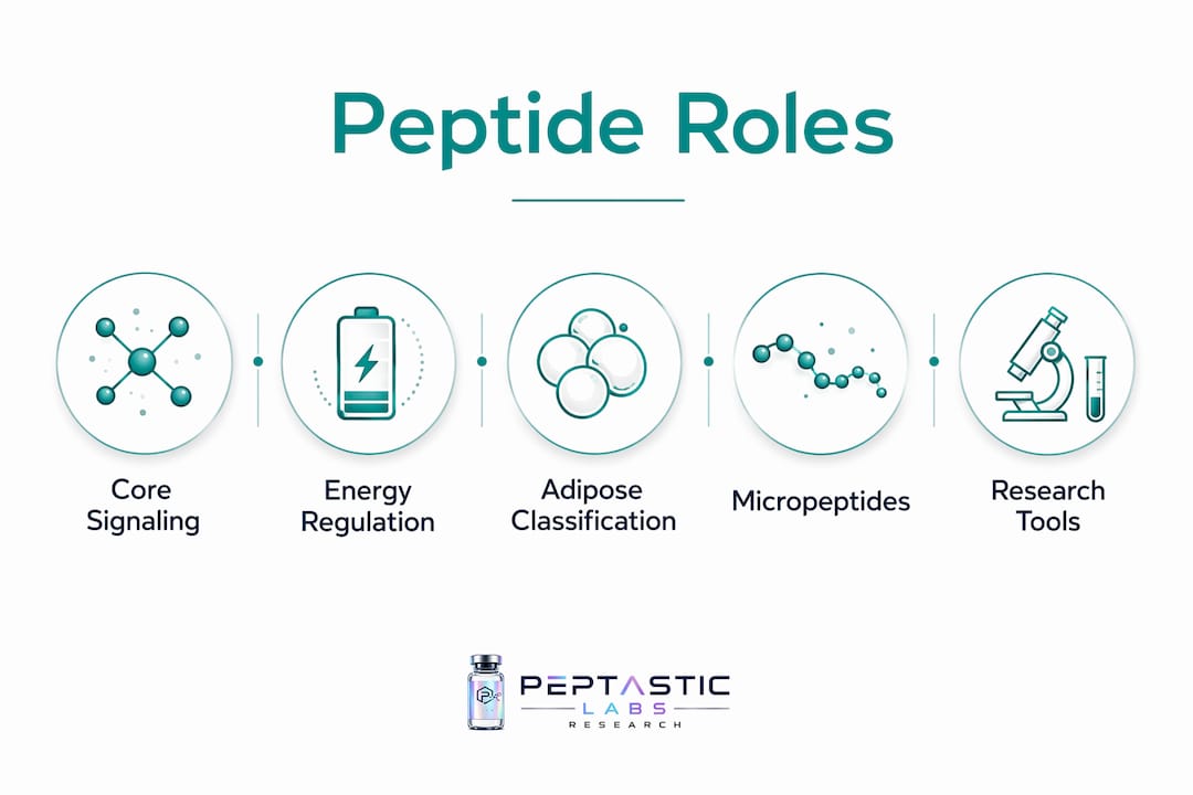
Peptides function as primary signaling molecules governing adipogenesis, lipolysis, energy expenditure, and inflammatory regulation within adipose tissue. The role of peptides in adipose tissue research spans classic adipokines such as leptin, adiponectin, and resistin, as well as a growing catalog of recently characterized peptide hormones and micropeptides. Their short half-lives and low toxicity profiles distinguish them from small-molecule drugs, making them attractive candidates for metabolic disease research. Key peptides and their primary functions in adipose biology include:
- Leptin: Signals fat mass to hypothalamic centers; regulates appetite and peripheral glucose metabolism
- Adiponectin: Enhances insulin sensitivity; activates AMPK; exerts anti-inflammatory effects
- Resistin: Pro-inflammatory adipokine; implicated in insulin resistance
- Adropin: Regulates energy homeostasis and lipid oxidation; expressed in adipose and liver tissue
- Apelin: Anti-inflammatory adipokine; modulates glucose uptake and adipogenesis
- Irisin: Secreted by muscle and adipose tissue; promotes white-to-beige fat conversion
- Kisspeptin: Regulates reproductive axis and energy balance via hypothalamic signaling
- MOTS-c: Mitochondria-derived peptide; activates AMPK; improves insulin sensitivity
- Phoenixin: Neuropeptide with roles in appetite suppression and reproductive regulation
- Spexin: Suppresses food intake; modulates lipid metabolism and adipocyte function
- Neuropeptides B and W: Regulate feeding behavior and energy balance via hypothalamic circuits
- LEAO and LDAMP1: Micropeptides encoded by small open reading frames in noncoding RNAs; reshape adipose plasticity and insulin sensitivity
Table of Contents
- How adipose tissue is classified and why it matters for peptide research
- What types of peptides are relevant to adipose tissue research?
- How do peptides signal within adipose tissue at the molecular level?
- What is the role of peptides in energy homeostasis and obesity?
- Recent research advances: micropeptides and emerging adipose signals
- Research outlook: where does adipose peptide biology go from here?
- Methodologies and experimental models used in peptide-adipose tissue research
- Peptasticlabs supports your adipose tissue peptide research
- Key Takeaways
How adipose tissue is classified and why it matters for peptide research
Adipose tissue is not a passive lipid depot. It is a metabolically active endocrine organ that secretes a diverse array of signaling molecules, including peptide hormones, cytokines, and exosomal cargo, that communicate with the liver, skeletal muscle, pancreas, brain, and vasculature. Recognizing this endocrine identity is foundational to interpreting how peptides interact with fat tissue at the molecular level.
Three functionally distinct adipose tissue types are recognized in current research:
- White adipose tissue (WAT): Primary site of energy storage as triacylglycerols; major source of leptin, adiponectin, resistin, and numerous other adipokines; subdivided into visceral (VAT) and subcutaneous (SAT) depots with distinct secretory profiles
- Brown adipose tissue (BAT): Specialized for non-shivering thermogenesis via uncoupling protein 1 (UCP1); secretes batokines including FGF21 and neuregulin-4; metabolically protective
- Beige (brite) adipose tissue: Inducible thermogenic cells within WAT depots; can be recruited by cold exposure, beta-adrenergic stimulation, or specific peptides such as irisin
Depot-specific differences carry direct mechanistic implications. Visceral fat is primarily responsible for metabolic disturbances in obesity, partly because VAT adipokine secretion differs substantially from SAT. VAT releases higher levels of pro-inflammatory mediators and contributes disproportionately to hepatic lipid flux. SAT, by contrast, tends to secrete more adiponectin relative to its mass.
The stromal vascular fraction (SVF) within adipose tissue adds another layer of complexity. Endothelial cells, preadipocytes, fibroblasts, pericytes, and resident immune cells all contribute to the adipose secretome. Macrophage infiltration in obese WAT shifts the local peptide environment toward pro-inflammatory profiles, with consequences for systemic insulin sensitivity. Proteomics studies have identified hundreds of secreted proteins from adipose tissue, with cell-type-specific origin (adipocytes versus immune cells) determining whether a given peptide exerts protective or pathological effects.
Pro Tip: When designing adipose tissue peptide experiments, specify the depot (VAT vs. SAT) and the cell fraction (whole tissue vs. isolated adipocytes vs. SVF) in your methods. Secretome composition differs substantially across these fractions, and conflating them is a common source of irreproducible results.
What types of peptides are relevant to adipose tissue research?
Peptides relevant to adipose biology span a wide biochemical range, from large glycoprotein adipokines to sub-100-amino-acid micropeptides translated from previously annotated noncoding RNAs. Understanding peptide types and uses across these classes is prerequisite to designing mechanistically sound experiments.
Biochemically, peptides are amino acid chains linked by peptide bonds, typically ranging from 2 to several hundred residues. In adipose tissue research, three major classes are operationally relevant:
- Classic adipokines: Leptin, adiponectin, resistin, visfatin, chemerin, apelin, omentin. Secreted via classical ER-Golgi pathways; act through defined cell-surface receptors; well-characterized in obesity models.
- Peptide hormones and neuropeptides: Adropin, irisin, kisspeptin, MOTS-c, phoenixin, spexin, neuropeptides B and W. Produced in adipose, muscle, or neuroendocrine tissues; exert systemic effects on energy balance and adipose function.
- Micropeptides from small open reading frames (smORFs): LEAO, LDAMP1, and related molecules translated from lncRNAs and previously annotated noncoding regions; validated by ribosome profiling, CRISPR, and mass spectrometry.
| Peptide class | Primary origin | Canonical function | Key challenge |
|---|---|---|---|
| Classic adipokines (leptin, adiponectin) | Adipocytes, SVF | Energy homeostasis, insulin sensitivity | Receptor promiscuity; obesity-induced dysregulation |
| Peptide hormones (irisin, adropin) | Muscle, adipose, liver | Fat browning, lipid oxidation | Short half-life; tissue-specific receptor expression |
| Neuropeptides (kisspeptin, spexin, NPB/W) | Hypothalamus, adipose | Appetite, reproductive axis | Blood-brain barrier penetration; pleiotropic effects |
| Mitochondria-derived (MOTS-c) | Mitochondrial genome | AMPK activation, glucose uptake | Intracellular delivery; stability |
| Micropeptides (LEAO, LDAMP1) | lncRNA smORFs | Adipose plasticity, insulin signaling | Detection sensitivity; functional validation |
| Intracellular peptides | Proteasomal processing | Insulin signaling, glucose uptake | Rapid degradation; mass spectrometry detection limits |
Two challenges cut across all classes. First, short half-life limits in vivo duration of action, requiring either frequent dosing or structural modification in therapeutic contexts. Second, receptor promiscuity means a single peptide may activate overlapping pathways in multiple tissues simultaneously, complicating attribution of adipose-specific effects.
Food-derived bioactive peptides present an additional translation bottleneck: rapid enzymatic degradation and unfavorable pharmacokinetics constrain their utility beyond in vitro systems, a constraint that applies equally to endogenous peptides used as exogenous research tools.
How do peptides signal within adipose tissue at the molecular level?
Peptide signaling in adipose tissue operates through three spatially distinct modes: endocrine (systemic circulation to distant organs), paracrine (local cell-to-cell communication within the tissue), and autocrine (self-signaling within the secreting cell). Adipose-derived peptides frequently engage all three simultaneously, which is why isolating adipose-specific effects in vivo requires careful experimental design that accounts for brain and gut axes.
Several well-characterized molecular pathways illustrate the range of peptide action:
- AMPK activation by adiponectin: Adiponectin binds AdipoR1 and AdipoR2 receptors, activating AMP-activated protein kinase. This increases fatty acid oxidation, suppresses hepatic gluconeogenesis, and reduces intracellular ceramide levels. In obese states, circulating high-molecular-weight (HMW) adiponectin declines due to ER stress-mediated retention by ERp44, impairing this pathway.
- Leptin hypothalamic signaling: Leptin binds ObRb receptors on hypothalamic neurons, activating JAK2-STAT3 signaling to suppress appetite and increase sympathetic outflow to adipose tissue. Leptin resistance in obesity uncouples this circuit, allowing continued energy intake despite elevated circulating leptin.
- Irisin-mediated fat browning: Irisin, cleaved from FNDC5, binds integrin receptors on white adipocytes and activates UCP1 expression, promoting thermogenic conversion to a beige phenotype.
- Apelin receptor (APJ) signaling: Apelin activates APJ, a GPCR, reducing adipogenesis and improving glucose uptake via PI3K-Akt pathways. Its anti-inflammatory properties partly involve suppression of NF-κB signaling.
- Intracellular peptide action on insulin signaling: Intracellular peptides identified in rat adipose tissue, including diazepam binding inhibitor fragments, modulate insulin-induced glucose uptake when added exogenously to adipocytes. Their levels shift with high-caloric diet exposure, linking proteasomal processing to metabolic state.
- ERp44-adiponectin interaction modulation: Synthetic IgM-derived peptides compete with endogenous adiponectin for ERp44 binding, releasing trapped HMW oligomers from the ER. This mechanism is mechanistically distinct from thiazolidinediones, which alter gene expression rather than protein-protein interactions.
- MOTS-c nuclear translocation: Under metabolic stress, MOTS-c translocates from mitochondria to the nucleus, where it regulates antioxidant gene expression and AMPK-dependent metabolic reprogramming.
- Spexin and lipid metabolism: Spexin acts via galanin receptors (GALR2/3) to suppress fatty acid uptake in adipocytes and reduce food intake via hypothalamic circuits.
- Kisspeptin-GPR54 axis: Kisspeptin signals through GPR54 to regulate gonadotropin-releasing hormone neurons, linking adipose energy status to reproductive function.
- Phoenixin and appetite regulation: Phoenixin acts via GPR173 to suppress food intake and modulate anxiety-related feeding behavior, with expression detected in adipose depots.
- Neuropeptides B and W: Both peptides bind NPBWR1 and NPBWR2 receptors in the hypothalamus, regulating feeding behavior and energy expenditure with depot-specific expression in adipose tissue.
Ribosome profiling and CRISPR-based validation have confirmed that many peptides previously assumed to be noncoding products are in fact translated and biologically active. Systemic effects of these peptides may involve multiple organs simultaneously, making adipose-specific mechanistic attribution a persistent methodological challenge.
Effects on adipogenesis and lipolysis deserve specific attention. Leptin suppresses lipogenesis and promotes lipolysis via cAMP-PKA pathways. Adiponectin reduces lipid accumulation by increasing fatty acid oxidation. Resistin, conversely, promotes lipid storage and impairs insulin-stimulated glucose uptake. Apelin and irisin both shift the adipogenic program toward thermogenic phenotypes, though through distinct receptor systems.

What is the role of peptides in energy homeostasis and obesity?
Peptide dysregulation is a defining feature of obesity pathophysiology, not merely a downstream consequence. As adipose tissue expands, the secretome shifts: leptin rises while adiponectin falls, and pro-inflammatory peptides from infiltrating macrophages amplify insulin resistance. This shift in adipose tissue secretion toward pro-inflammatory cytokines creates a self-reinforcing cycle that worsens metabolic dysfunction.
Key peptide-level changes in obesity include:
- Leptin resistance: Circulating leptin is elevated in obesity, but hypothalamic ObRb signaling is blunted, decoupling the adipostat signal from appetite suppression
- Reduced HMW adiponectin: ER stress in obese adipocytes traps adiponectin via ERp44, reducing circulating HMW fractions and impairing AMPK-mediated insulin sensitization
- Elevated resistin: Promotes hepatic insulin resistance and systemic inflammation; secreted by macrophages within obese WAT in humans
- Suppressed apelin: Circulating apelin declines with increasing adiposity, removing an anti-inflammatory and insulin-sensitizing signal
- Altered irisin: Exercise-induced irisin secretion is blunted in sedentary obese states, reducing thermogenic recruitment of beige adipocytes
- Spexin deficiency: Plasma spexin levels are consistently lower in obese individuals compared to lean controls, correlating with impaired lipid metabolism
The therapeutic implications of these shifts have driven considerable research interest, with a range of peptides Australia weight loss alternatives being explored for metabolic modulation. Synthetic IgM-derived peptides targeting the ERp44-adiponectin interaction increased circulating adiponectin and reduced triglycerides and cholesterol in high-fat diet mouse models, without altering adiponectin gene expression. This mechanistic distinction from thiazolidinediones is significant: the peptide approach modulates protein assembly rather than transcription, potentially avoiding the gene-expression-level side effects associated with PPARγ agonists.
Adipose tissue peptide research also intersects with immune modulation. Macrophage infiltration in obese WAT generates a pro-inflammatory peptide environment that accelerates adipocyte dysfunction. Peptides with immune-modulating properties such as adiponectin and apelin counterbalance this shift, but their concentrations decline precisely when they are most needed.
Proteomics studies have identified hundreds of secreted proteins from adipose tissue, with depot-specific variation and cell-type-specific origin determining whether a given peptide exerts protective or pathological effects on systemic metabolism.
The complexity of the adipose secretome means no single peptide fully explains obesity-associated metabolic dysfunction. MOTS-c, for instance, improves insulin sensitivity and activates AMPK independently of leptin or adiponectin pathways, suggesting that mitochondria-derived signals contribute to adipose energy regulation through mechanisms that remain incompletely characterized.
Recent research advances: micropeptides and emerging adipose signals
The most consequential shift in adipose peptide research over the past decade has been the recognition that the noncoding genome encodes functional peptides. Small open reading frames (smORFs) within long noncoding RNAs (lncRNAs) and other previously annotated noncoding transcripts produce micropeptides that regulate lipid storage, adipose plasticity, and insulin sensitivity in ways that classical adipokine biology did not anticipate.
Key recent findings include:
- LEAO: A micropeptide encoded by a smORF within a lncRNA; reshapes adipose tissue plasticity and improves metabolic homeostasis in obesity models; validated by genome editing and mass spectrometry
- LDAMP1: Regulates lipid storage and insulin sensitivity; identified through ribosome profiling and CRISPR-based functional screens
- Intracellular proteasomal peptides: Mass spectrometry has detected adipose-derived intracellular peptides whose exogenous addition to adipocytes increases glucose uptake, with levels altered by high-caloric diet exposure
- Exosomal peptide cargo: FABP4 and other adipocyte-derived proteins secreted via exosomes function as circulating adipokines, expanding the secretome beyond classically secreted proteins
Adipose tissue plasticity is dynamic, and its loss during obesity is pathological. Newly discovered micropeptides expand the mechanistic understanding of this plasticity and open research directions that classical adipokine biology could not address.
The methodological advances enabling these discoveries are inseparable from the findings themselves. Ribosome profiling (Ribo-seq) identifies actively translated smORFs genome-wide. CRISPR-Cas9 knockout and knockin approaches validate functional necessity. High-resolution mass spectrometry, particularly semiquantitative LC-MS/MS, detects low-abundance micropeptides that Western blot cannot resolve. These tools together have made it feasible to characterize the full translational output of adipose tissue, not just the proteins predicted by canonical annotation.
Challenges in this space remain substantial. Peptide signaling promiscuity means that a micropeptide validated in one adipose depot or species may act through entirely different receptors in another context. Complex crosstalk between adipose peptide pathways and gut, liver, and brain signals makes adipose-specific attribution difficult even with tissue-specific knockout models. Translating micropeptide findings from rodent models to human adipose biology requires careful attention to species-specific lncRNA sequences, which often lack direct orthologs.
Recent advances in this area are organized below by discovery approach:
- Ribosome profiling-validated smORF peptides: LEAO, LDAMP1, and related molecules confirmed as translated products of lncRNA loci
- Proteomics-identified secretome expansions: Hundreds of novel adipokines identified via LC-MS/MS from primary adipocyte cultures and adipose tissue explants
- Exosome-associated peptide signals: FABP4 and exosomal miRNA-peptide complexes identified as non-classical secretory products with systemic metabolic effects
- Mitochondria-derived peptides: MOTS-c and humanin characterized as mitochondrial genome-encoded regulators of AMPK and insulin signaling in adipose tissue
- Diet-responsive intracellular peptides: Proteasomal processing products in rat adipose tissue whose abundance shifts with caloric intake, linking energy status to intracellular signaling peptide pools
Research outlook: where does adipose peptide biology go from here?
The field has moved well past cataloging adipokines. Current priorities center on mechanistic resolution, translational validation, and therapeutic specificity.
Key summary points from the current state of adipose peptide research:
- Adipose tissue secretes a complex mixture of peptides from adipocytes, immune cells, endothelial cells, and preadipocytes; cell-type-specific origin determines functional outcome
- Classic adipokines (leptin, adiponectin, resistin) regulate energy homeostasis, insulin sensitivity, and inflammation through well-defined receptor systems, but their dysregulation in obesity is multifactorial
- Peptide hormones (adropin, apelin, irisin, kisspeptin, MOTS-c, phoenixin, spexin, neuropeptides B and W) extend adipose signaling into thermogenesis, reproductive regulation, and mitochondrial metabolism
- Micropeptides from smORFs represent a newly validated layer of adipose regulation with direct relevance to fat browning and insulin signaling
- Therapeutic targeting of peptide pathways, as demonstrated by ERp44-adiponectin interaction modulators, can improve metabolic parameters without altering gene expression profiles
Gaps that warrant priority attention include:
- Human validation of micropeptide findings currently established only in rodent models
- Depot-specific and cell-type-specific secretome characterization using single-cell proteomics
- Pharmacokinetic stabilization strategies for short-half-life peptides intended for in vivo use
- Mechanistic dissection of receptor promiscuity to enable tissue-targeted peptide design
- Integration of multi-omics data (transcriptomics, proteomics, metabolomics) to map the full adipose peptide signaling network
The mTOR pathway represents one underexplored node in this network. Researchers investigating mTOR pathway peptide interactions in adipose tissue will find that mTORC1 and mTORC2 both intersect with adipokine secretion and adipogenesis, offering additional mechanistic entry points for peptide-based intervention.
Multi-omics integration is not optional at this stage. Single-cell RNA sequencing has already revealed that adipose tissue contains far more cell-type diversity than bulk transcriptomics suggested. Applying analogous resolution to the proteome and secretome will be necessary to assign peptide functions to specific cell populations rather than to whole-tissue averages.
Methodologies and experimental models used in peptide-adipose tissue research
Methodological rigor determines whether findings in this field replicate or collapse. The complexity of adipose tissue biology demands matched experimental systems.
In vitro models
3T3-L1 murine preadipocytes remain the most widely used cell line for adipogenesis and peptide-response studies. Primary human preadipocytes from subcutaneous or visceral depots offer greater translational relevance but introduce donor variability. Adipose tissue explants preserve the multicellular environment, including SVF contributions, and are preferable for secretome studies over isolated adipocyte monocultures.
For peptide detection, semiquantitative LC-MS/MS outperforms Western blot for low-abundance and small peptides. Western blot is prone to cross-reactivity artifacts with micropeptides below 10 kDa, and its dynamic range is insufficient for detecting subtle concentration changes in conditioned media. ELISA remains appropriate for well-characterized adipokines (leptin, adiponectin) with validated antibodies, but should not be the primary detection method for novel peptides.
In vivo models
Diet-induced obesity (DIO) models using high-fat diet in C57BL/6 mice are standard for studying obesity-associated peptide dysregulation. Genetic models (ob/ob, db/db) provide leptin-deficient or leptin-receptor-deficient backgrounds useful for dissecting leptin-independent peptide pathways. Tissue-specific knockout models, generated via Cre-lox recombination with adiponectin-Cre or Fabp4-Cre drivers, allow depot-specific and cell-type-specific peptide deletion without systemic confounds.

Careful experimental design is required to differentiate autocrine and paracrine peptide actions from endocrine effects. Conditioned medium transfer experiments, parabiosis models, and tissue-specific peptide overexpression each address different aspects of this question. Systemic peptide infusion studies must account for brain and gut axis effects that may confound adipose-specific interpretations.
Proteomic and genomic tools
Ribosome profiling (Ribo-seq) is now standard for identifying translated smORFs in adipose tissue transcriptomes. CRISPR-Cas9 provides functional validation of candidate micropeptides through targeted deletion or tagging. Proximity labeling approaches (BioID, TurboID) map peptide interactomes within adipocytes without requiring stable overexpression of large fusion proteins.
For synthetic peptide studies, verifying cell penetration and intracellular stability is prerequisite. Cell-penetrating peptide (CPP) conjugation strategies, as used in ERp44-adiponectin research, require confirmation that the CPP sequence does not independently alter the metabolic readout being measured. Dose-response and time-course experiments should be conducted before interpreting any single-concentration result.
Pro Tip: When validating a newly identified adipose micropeptide, combine Ribo-seq with CRISPR knockout and mass spectrometry-based quantification in at least two independent cell systems before advancing to in vivo models. Single-method validation of smORF-encoded peptides has a high false-positive rate.
Peptasticlabs supports your adipose tissue peptide research
Researchers working at the intersection of peptide biology and metabolic disease need compounds they can trust before the experiment begins. Peptasticlabs supplies research-grade peptides verified to ≥99% purity via HPLC, with third-party testing and batch-specific Certificates of Analysis available on request. Every compound in the catalog undergoes in-house quality checks before third-party verification, eliminating the sourcing uncertainty that compromises reproducibility.

The catalog covers peptides directly relevant to adipose tissue and metabolic research, including compounds targeting AMPK activation, insulin signaling, and thermogenic pathways. For researchers studying adipokine interactions, ERp44-related mechanisms, or micropeptide functional validation, compound purity and documentation are not secondary concerns. Peptasticlabs structures its entire supply chain around that requirement. Certificates of Analysis are provided per batch, not per product line, so you know exactly what went into your experiment. Browse the full research-grade peptide catalog to identify compounds matched to your current protocol.
Key Takeaways
Peptides regulate adipose tissue function through endocrine, paracrine, and autocrine signaling pathways that govern adipogenesis, lipolysis, energy homeostasis, and inflammation, with obesity disrupting these circuits at multiple molecular levels.
| Point | Details |
|---|---|
| Adipose tissue is an endocrine organ | WAT, BAT, and beige depots each secrete distinct peptide profiles that regulate systemic metabolism and inflammation. |
| Obesity shifts the peptide secretome | Leptin rises, HMW adiponectin declines, and pro-inflammatory macrophage-derived peptides increase, worsening insulin resistance. |
| Micropeptides expand the field | LEAO and LDAMP1, encoded by smORFs in lncRNAs, regulate adipose plasticity and insulin sensitivity in ways classical adipokines do not. |
| Synthetic peptides show therapeutic specificity | IgM-derived peptides targeting ERp44-adiponectin interactions improved insulin sensitivity and reduced lipids in obese mice without altering gene expression. |
| Peptasticlabs for research use | HPLC-verified, ≥99% purity compounds with batch-specific Certificates of Analysis support reproducible adipose peptide research. |