
Glucose-dependent insulinotropic polypeptide (GIP) is a 42-amino acid incretin hormone secreted primarily from upper intestinal K cells that potentiates glucose-stimulated insulin secretion and coordinates postprandial nutrient storage across multiple tissues. Understanding the full function of GIP peptide requires moving well beyond its pancreatic role: GIPR is expressed in adipose tissue, bone, adrenal cortex, and multiple brain regions, making GIP a pleiotropic metabolic regulator with direct translational implications.
- Canonical incretin action: GIP amplifies glucose-dependent insulin secretion from pancreatic β-cells, accounting for a substantial portion of the postprandial insulin response. This incretin effect is absent with intravenous glucose, which is the defining experimental signature.
- Extrapancreatic targets: GIP acts directly on white and brown adipose tissue (stimulating lipogenesis and inhibiting lipolysis), on bone (promoting osteoblast proliferation and suppressing osteoclastic resorption), and on CNS nuclei including the hypothalamus and brainstem, where it modulates appetite and nausea.
- Translational relevance: Native GIP is rapidly inactivated by DPP-4, complicating both measurement and therapeutic use. The GIP/GLP-1 co-agonist tirzepatide has demonstrated superior weight loss and glycemic control versus GLP-1 monoagonists in randomized trials, supporting dual incretin targeting as a promising approach.
Key Takeaways
GIP is a pleiotropic incretin with glucose-dependent insulinotropic action, direct effects on adipose tissue, bone, and CNS, and a therapeutic paradox that makes both GIPR agonism and antagonism metabolically relevant depending on context.
| Point | Details |
|---|---|
| GIP is a 42-amino acid incretin | GIP(1–42) potentiates glucose-dependent insulin secretion from pancreatic β-cells postprandially. |
| DPP-4 inactivation is rapid | Native GIP has a plasma half-life of approximately 5–7 minutes; DPP-4 inhibition in collection tubes is required for accurate active GIP measurement. |
| Extrapancreatic roles are substantial | GIPR drives lipogenesis in adipose tissue, osteoblast activity in bone, and appetite/nausea modulation in brainstem nuclei. |
| The obesity paradox is unresolved | Endogenous GIP elevation correlates with adiposity, yet GIP/GLP-1 co-agonism produces superior weight loss versus GLP-1 monoagonism in RCTs. |
| Peptasticlabs for reagent QC | Peptasticlabs supplies HPLC-verified, ≥99% purity peptides with batch-specific CoA and third-party testing for reproducible GIP research. |
Table of Contents
- How GIP peptide function begins: biosynthesis, K-cell secretion, and nutrient triggers
- Where GIPR is expressed and how receptor signaling drives tissue responses
- GIP's insulinotropic actions and how they compare to GLP-1
- Extrapancreatic roles of GIP across adipose tissue, bone, CNS, and other organs
- DPP-4 degradation, plasma half-life, and assay best practices for measuring GIP
- The GIP-obesity paradox: why endogenous elevation and therapeutic agonism produce opposite outcomes
- Translational and clinical evidence for GIP/GLP-1 co-agonists
- Practical methods for researchers measuring and manipulating GIP
- Conclusions: GIP's physiological role and the research questions that remain
- The GIP paradox deserves more attention than it gets
- Sourcing research-grade GIP reagents: what to verify before you order
- Sources
How GIP peptide function begins: biosynthesis, K-cell secretion, and nutrient triggers
GIP is synthesized as a 153-amino acid preproGIP precursor. Tissue-specific post-translational processing cleaves preproGIP to proGIP, then to the biologically active GIP(1–42), the 42-amino acid form that circulates after a meal. A truncated form, GIP(1–30)NH₂, is produced in some tissues but GIP(1–42) is the dominant circulating species in humans.
The primary source of GIP is the K cell, an enteroendocrine cell concentrated in the duodenum and proximal jejunum. Historically, the peptide was named gastric inhibitory polypeptide based on early observations of gastric acid suppression. That inhibitory effect on acid secretion is negligible in humans at physiological concentrations, and the field now uses glucose-dependent insulinotropic polypeptide to reflect its actual primary function. Researchers should use the modern nomenclature to avoid ambiguity in literature searches and protocol descriptions.
Three macronutrient classes drive K-cell secretion, but their mechanisms differ:
- Oral glucose: Absorbed via SGLT1 in the proximal intestine; luminal glucose is the most studied GIP secretagogue. Critically, intravenous glucose does not stimulate GIP release, which is the basis of the incretin effect paradigm.
- Dietary fat: Long-chain fatty acids activate free fatty acid receptor 1 (FFAR1/GPR40) and related GPCRs on K cells; fat is a potent GIP secretagogue and produces a prolonged postprandial GIP elevation.
- Protein and peptides: Protein hydrolysates stimulate GIP, though the receptor mechanisms are less characterized than for glucose or fat.
For study design, macronutrient composition matters substantially. A high-fat meal produces a different GIP kinetic profile than an isocaloric glucose load, and mixed meals generate the highest total GIP exposure. Postprandial GIP peaks within 15–30 minutes of nutrient ingestion and returns toward baseline within 2–3 hours, though this window shifts with meal composition and individual variation. Sampling at a single postprandial time point will miss the full secretory arc.
Pro Tip: When designing a GIP secretion study, collect samples at 0, 15, 30, 60, 90, and 120 minutes postprandially. A single 60-minute draw is insufficient to characterize peak secretion, particularly after high-fat meals where the GIP curve is broader and later-peaking than after a glucose load.
Where GIPR is expressed and how receptor signaling drives tissue responses
The GIP receptor (GIPR) is a class B G protein-coupled receptor with broad tissue distribution that explains GIP's pleiotropic effects. Confirmed GIPR expression sites include:
- Pancreatic β-cells (primary incretin target)
- White and brown adipose tissue
- Cortical and trabecular bone (osteoblasts and osteoclasts)
- Adrenal cortex
- Heart and vascular endothelium
- Anterior pituitary
- Hypothalamus, area postrema, nucleus tractus solitarius (NTS), and other brainstem nuclei
- Peripheral nervous system ganglia
At the cellular level, GIPR couples predominantly to Gαs, activating adenylate cyclase and raising intracellular cAMP. Elevated cAMP activates both protein kinase A (PKA) and the exchange protein directly activated by cAMP 2 (Epac2). In β-cells, PKA phosphorylates components of the exocytotic machinery and closes KATP channels, while Epac2 facilitates Ca²⁺ mobilization and granule priming. Phospholipase C (PLC) and protein kinase C (PKC) contributions have been described in some tissues, though these are secondary to the cAMP axis in most contexts. Biased signaling at GIPR, where different ligands preferentially activate distinct downstream pathways, is an active area of investigation with direct implications for therapeutic design.
Receptor distribution studies rely on immunohistochemistry (IHC), radioligand autoradiography, and mRNA expression profiling. Each method carries caveats. IHC antibody specificity for GIPR has been inconsistent across commercial sources, and several published distribution maps used reagents later shown to have off-target binding. Autoradiography with radiolabeled GIP analogues provides functional binding data but cannot distinguish receptor subtypes. mRNA expression confirms transcription but not surface protein levels. When extrapolating animal receptor distribution data to humans, note that rodent GIPR expression in the CNS is more extensively characterized than human CNS expression, and direct human mapping remains incomplete.
GIP's insulinotropic actions and how they compare to GLP-1
GIP potentiates glucose-stimulated insulin secretion in a strictly glucose-dependent manner: at fasting glucose concentrations, GIP has minimal insulinotropic effect, but as plasma glucose rises postprandially, GIP amplifies β-cell insulin output substantially. This glucose dependency is a safety feature that limits hypoglycemia risk and distinguishes incretins from sulfonylureas.
The incretin effect is defined as the difference in insulin secretion following oral versus isocaloric intravenous glucose. GIP and GLP-1 together account for the majority of this effect, with GIP historically credited with a larger share under standard mixed-meal conditions. GIP(1–42) contributes substantially to the postprandial incretin effect and is rapidly degraded by DPP-4, which shortens its circulating half-life and limits the duration of its insulinotropic signal. For researchers designing incretin studies, the oral versus IV glucose clamp comparison remains the gold-standard protocol for quantifying the incretin effect and isolating GIP's contribution.
Statistic callout: Studies using the oral glucose infusion method to match plasma glucose profiles between oral and IV routes have demonstrated that the incretin effect accounts for more than half of total postprandial insulin secretion in healthy individuals, with GIP and GLP-1 thought to be the primary mediators.
GIP and GLP-1 share the incretin mechanism but diverge in several physiologically important ways. GLP-1 suppresses glucagon secretion from α-cells; GIP stimulates glucagon under hypoglycemic conditions, which may serve a counterregulatory function. GLP-1 slows gastric emptying; GIP has no consistent effect on gastric motility in humans. GLP-1 receptor agonists produce dose-dependent nausea via area postrema activation; GIP, acting through brainstem GIPR, can attenuate this nausea response. These differences explain why GIP alone failed to produce meaningful glycemic benefit in early type 2 diabetes trials (likely due to GIPR desensitization or downregulation in the insulin-resistant state) while the GIP/GLP-1 combination has demonstrated additive or synergistic metabolic effects.
Extrapancreatic roles of GIP across adipose tissue, bone, CNS, and other organs
GIP's anabolic effects extend well beyond the pancreas, with direct receptor-mediated actions documented in multiple tissues. The evidence base varies considerably by tissue and species.
Adipose tissue
GIP stimulates lipoprotein lipase (LPL) activity in adipocytes, facilitating fatty acid uptake from circulating triglycerides. It promotes lipogenesis and glycogen synthesis while inhibiting hormone-sensitive lipase, effectively suppressing lipolysis. These actions position GIP as a postprandial nutrient-storage signal in fat tissue. In obesity, circulating GIP is chronically elevated and GIPR expression in adipose tissue may be altered, contributing to the adiposity-GIP feedback loop discussed in Section 7. Evidence: primarily human and rodent in vitro and in vivo data.
Bone
GIPR is expressed on osteoblasts and osteoclasts. GIP stimulates osteoblast proliferation and collagen synthesis while inhibiting osteoclastic bone resorption, producing a net anabolic effect on bone. Postprandial GIP spikes may contribute to the "meal-related bone remodeling" phenomenon observed in humans. The clinical implication is that GIP-targeting therapies could have off-target effects on bone mineral density, a parameter worth including in longer-term metabolic trials. Evidence: human and animal data, with some human observational support.
CNS: appetite, satiety, and nausea
GIPR expression in the hypothalamus, area postrema, and NTS positions GIP as a central regulator of energy balance. CNS GIPR agonism recruits GABAergic circuits in the area postrema and NTS that suppress appetite and, critically, attenuate GLP-1-related nausea. This mechanism is central to the tolerability advantage of GIP/GLP-1 co-agonists. Evidence: primarily rodent mechanistic data with translational support from co-agonist clinical trials.

GI tract, kidney, and heart
GIP's original namesake function, gastric acid inhibition, is not physiologically relevant at normal postprandial concentrations in humans. Kidney clearance contributes to GIP elimination alongside DPP-4 degradation. Cardiac GIPR expression has been documented, and some preclinical data suggest cardioprotective signaling, though human cardiovascular evidence for GIP specifically remains limited.
| Tissue | Primary GIP Effect | Direction | Evidence Strength |
|---|---|---|---|
| Pancreatic β-cell | Insulin secretion potentiation | Increase | Strong (human RCT) |
| White adipose | LPL activity, lipogenesis, lipolysis | Increase/Decrease | Moderate (human + animal) |
| Bone | Osteoblast activity, osteoclast resorption | Increase/Decrease | Moderate (human observational + animal) |
| Hypothalamus/brainstem | Appetite suppression, nausea attenuation | Decrease | Moderate (animal + translational) |
| Stomach (acid) | Gastric acid secretion | Negligible in humans | Weak (historical, not replicated) |
| Heart | Cardioprotective signaling | Increase (preclinical) | Preliminary (animal only) |
DPP-4 degradation, plasma half-life, and assay best practices for measuring GIP
Native GIP is rapidly inactivated by DPP-4, which cleaves the N-terminal His-Ala dipeptide to produce GIP(3–42) DPP-4 processing and incretin stability study - PubMed. This truncated metabolite has markedly reduced insulinotropic activity and may act as a partial antagonist at GIPR in some contexts. The plasma half-life of intact GIP(1–42) is typically reported in the range of 5–7 minutes in humans, making ex vivo degradation a significant source of measurement error if samples are not handled correctly.
For researchers, the practical implications are substantial. The following numbered protocol addresses the most common assay failures:
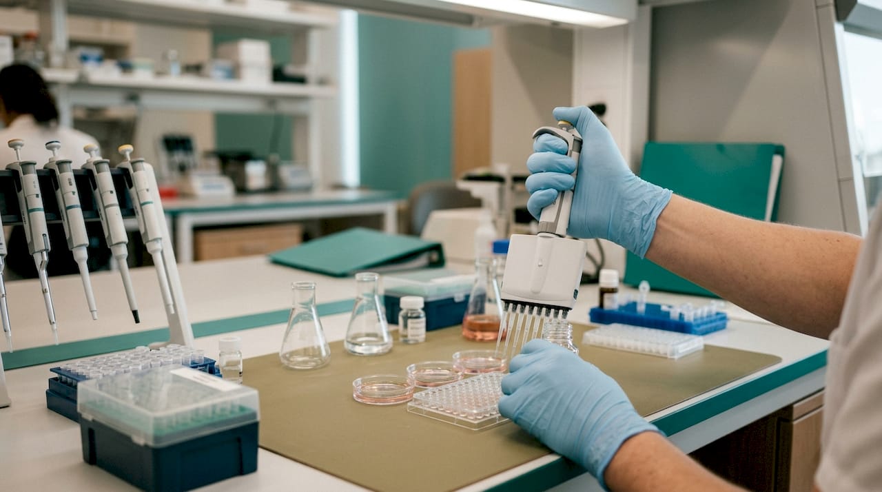
- Collection tubes: Use EDTA-plasma tubes pre-loaded with a DPP-4 inhibitor (e.g., diprotin A or a commercially validated DPP-4 inhibitor cocktail). Plain serum tubes allow rapid ex vivo cleavage and will underestimate active GIP.
- Temperature: Place tubes on ice immediately after collection. Do not leave at room temperature.
- Processing time: Centrifuge within 30 minutes of collection. Delays beyond 60 minutes at ambient temperature substantially reduce measured active GIP.
- Aliquoting and storage: Aliquot plasma into low-binding tubes and store at -80°C. Avoid repeated freeze-thaw cycles; each cycle degrades active GIP further.
- Assay selection: Immunoassays (ELISA, RIA) are widely used but antibody specificity is critical. Antibodies that recognize the C-terminus measure total GIP (active + inactive). N-terminal or mid-region antibodies may preferentially detect active GIP(1–42). Confirm cross-reactivity with GIP(3–42) for your specific assay.
- LC-MS confirmation: Liquid chromatography-mass spectrometry can differentiate GIP(1–42) from GIP(3–42) and other metabolites with high specificity. Use LC-MS as the reference method when validating a new immunoassay or when active versus total GIP discrimination is critical to the study question.
- Reporting: State explicitly whether your assay measures total or active GIP. Many older studies used total GIP assays without DPP-4 inhibition, making cross-study comparisons unreliable.
The distinction between total and active GIP is not academic. A study reporting elevated postprandial GIP in a patient group may be detecting accumulated inactive GIP(3–42) rather than a true increase in insulinotropic signal. Boosting GIPR signaling in combination with GLP-1 has shown metabolic benefits in recent translational work, but interpreting those findings requires knowing whether the measured GIP reflects the active ligand.
The GIP-obesity paradox: why endogenous elevation and therapeutic agonism produce opposite outcomes
Endogenous GIP overexpression correlates with adiposity and insulin resistance, yet controlled GIPR agonism combined with GLP-1 receptor activation produces clinically meaningful weight loss in randomized trials. This apparent contradiction is one of the most discussed mechanistic puzzles in incretin biology.
The paradox may resolve around context: chronic, uncontrolled GIP elevation in obesity drives adipose nutrient deposition and may desensitize GIPR in peripheral tissues, while acute, pharmacological GIPR agonism at CNS sites, particularly the hypothalamus and brainstem, engages appetite-suppressing and nausea-attenuating circuits that dominate the metabolic outcome when GLP-1 co-agonism is present. The therapeutic effect is not simply "more GIP" but rather a qualitatively different receptor engagement pattern, in a different ligand and dosing context, at sites that are not chronically desensitized.
Several mechanistic hypotheses have been proposed:
- Receptor desensitization: Chronic GIP elevation in obesity may downregulate or desensitize peripheral GIPR (particularly in adipose and β-cells), blunting the insulinotropic response while leaving CNS GIPR more responsive to pharmacological agonism.
- CNS versus peripheral balance: Peripheral GIPR activation promotes nutrient storage; central GIPR activation suppresses intake. In obesity, the peripheral signal dominates endogenously, but pharmacological co-agonists may shift the balance toward central effects.
- Ligand bias: Stabilized GIP analogues used therapeutically may engage GIPR signaling pathways differently than native GIP(1–42), activating β-arrestin or cAMP pathways in tissue-specific ratios that differ from the endogenous ligand.
- GLP-1 co-engagement: GIP's metabolic benefits in trials may be partially or largely dependent on simultaneous GLP-1 receptor activation, with the two systems producing synergistic effects that neither achieves alone.
Preclinical genetic ablation and immunoneutralization of GIP signaling reduce obesity in rodent models, supporting a causal role for GIP in adiposity. Yet the same models show that GIPR agonism can reduce food intake when administered centrally. These findings are not mutually exclusive: GIP promotes fat storage peripherally and can suppress appetite centrally, and which effect dominates depends on the experimental or therapeutic context.
Caveats are substantial. Rodent GIPR pharmacology does not map cleanly to humans. Dosing regimens, receptor occupancy, and the relative contribution of central versus peripheral GIPR engagement differ between species and between native peptide and stabilized analogues. Long-term human data on GIPR modulation beyond glycemia and weight, particularly on bone, cardiovascular endpoints, and CNS function, remain limited.
Translational and clinical evidence for GIP/GLP-1 co-agonists
The GIP/GLP-1 co-agonist tirzepatide has produced greater reductions in HbA1c and body weight than GLP-1 receptor monoagonists in head-to-head randomized controlled trials, establishing the clinical proof-of-concept for dual incretin targeting. The magnitude of weight loss observed with tirzepatide in the SURMOUNT program exceeded what had been seen with GLP-1 monoagonists at comparable timepoints, though direct mechanistic attribution to the GIP component versus the GLP-1 component remains an active research question.
Key trial findings from published randomized data:
- Glycemic control: HbA1c reductions with GIP/GLP-1 co-agonism exceeded those with GLP-1 monoagonism across multiple dose comparisons in type 2 diabetes populations.
- Weight loss: Co-agonist treatment produced body weight reductions in the range of 15–22% from baseline in obesity trials, with the highest doses approaching outcomes previously seen only with bariatric surgery.
- Tolerability: Gastrointestinal adverse events (nausea, vomiting, diarrhea) occurred at rates broadly comparable to GLP-1 monoagonists, with some trial data suggesting a more favorable nausea profile for co-agonists, consistent with the CNS GIPR mechanism described above.
- Cardiovascular signals: Early cardiovascular outcome data are emerging; longer-term dedicated trials are ongoing.
For researchers designing studies on GIP-targeting agents, recommended endpoints beyond glycemia and weight include: fasting and postprandial GIP (active, not total), GLP-1, insulin, C-peptide, glucagon, bone turnover markers (P1NP, CTX), appetite-related scores, and nausea/GI symptom diaries. Including CNS-targeted biomarkers (e.g., functional MRI appetite paradigms in human studies) would substantially advance mechanistic understanding.
Comparison: GIP/GLP-1 co-agonist versus GLP-1 monoagonist
| Parameter | GIP/GLP-1 co-agonist | GLP-1 monoagonist |
|---|---|---|
| HbA1c reduction | Greater (head-to-head RCT data) | Clinically significant |
| Body weight reduction | Greater; up to 22% in obesity trials | Up to ~15% at highest doses |
| Nausea/GI tolerability | Comparable or modestly favorable | Dose-dependent nausea common |
| Bone effects | Under investigation | Minimal data |
| CNS appetite mechanism | GIPR + GLP-1R engagement | GLP-1R-mediated |
| Glucagon response | GIP stimulates glucagon under hypoglycemia | GLP-1 suppresses glucagon |
Practical methods for researchers measuring and manipulating GIP
Rigorous GIP research requires attention to sample handling, assay selection, and peptide reagent quality at every stage. The following protocol checklist is designed for direct use in study planning.
Sample handling protocol
- Pre-load collection tubes with a validated DPP-4 inhibitor before the study visit.
- Collect blood into EDTA tubes; place immediately on ice.
- Centrifuge at 4°C within 30 minutes of collection.
- Aliquot plasma into low-binding polypropylene tubes; label with time point and condition.
- Store at -80°C; limit freeze-thaw cycles to one per aliquot.
- Document collection-to-centrifuge time for every sample; exclude samples with delays exceeding 60 minutes.
Assay selection
- Immunoassay (ELISA/RIA): High throughput, widely available, lower cost. Antibody specificity is the critical variable. Confirm whether the assay detects GIP(1–42) only, total GIP, or both. Request the manufacturer's cross-reactivity data for GIP(3–42) before committing to a platform.
- LC-MS: Reference-standard specificity; differentiates active from inactive forms. Higher cost and throughput constraints make it impractical for large cohorts but appropriate for method validation and mechanistic studies.
- Active versus total GIP: For most translational studies, active GIP (with DPP-4 inhibition in the tube) is the relevant measurement. Total GIP assays are acceptable for pharmacokinetic studies of stabilized analogues where the analogue itself is the target.
Peptide reagent quality checklist
When sourcing GIP peptide or GIP analogues for in vitro or in vivo research, verify the following before ordering:
- Purity: ≥99% by HPLC; request the HPLC trace, not just the stated value.
- Certificate of Analysis (CoA): Batch-specific, not a generic template. Should include molecular weight confirmation, purity, and sequence verification.
- Third-party testing: Independent verification of purity and identity, separate from the supplier's in-house QC.
- Storage and shipping: Lyophilized peptides shipped with cold packs; storage at -20°C or -80°C depending on stability data.
- Reconstitution guidance: Supplier should specify the recommended solvent (sterile water, acetic acid, DMSO) and concentration range to avoid aggregation.
Animal dosing and CNS targeting
For rodent studies, peripheral GIP administration (subcutaneous or intravenous) does not reliably engage CNS GIPR at the same receptor occupancy as intracerebroventricular (ICV) injection. If the research question involves central appetite or nausea mechanisms, ICV or direct nucleus-targeted delivery is required to isolate CNS effects from peripheral ones. Peripheral dosing studies that report "central effects" without CNS delivery confirmation should be interpreted cautiously. For metabolic peptide study design, including appropriate vehicle controls and pair-feeding controls is standard practice when energy intake is an endpoint.
Pro Tip: Always run a parallel sample set with and without DPP-4 inhibitor in your first GIP assay validation experiment. The ratio of active to total GIP in your specific matrix and handling conditions will tell you whether your standard protocol is adequate or whether you need stricter temperature control.
Conclusions: GIP's physiological role and the research questions that remain
Three statements summarize the current state of GIP peptide function:
- GIP is a glucose-dependent insulinotropic incretin secreted postprandially from duodenal K cells, with a primary role in amplifying β-cell insulin output and coordinating nutrient storage across adipose tissue and bone.
- GIPR's broad tissue distribution, from pancreas to brainstem, produces pleiotropic metabolic effects that extend well beyond insulin secretion and that are only partially characterized in humans.
- Rapid DPP-4 inactivation limits the circulating half-life of native GIP and creates systematic measurement artifacts in studies that do not use DPP-4 inhibition during sample collection.
The most tractable open research questions are:
- CNS versus peripheral GIPR mapping in humans: Rodent data on hypothalamic and brainstem GIPR are extensive; human CNS receptor distribution and functional engagement during pharmacological GIPR agonism remain poorly characterized. PET ligand development and human neuroimaging studies are needed.
- Long-term GIPR modulation and bone: GIP's anabolic bone effects are documented in short-term studies, but the long-term skeletal consequences of sustained GIPR agonism or antagonism in metabolic disease populations have not been systematically evaluated.
- Assay harmonization across clinical trials: Active GIP measurement protocols vary substantially across published trials, making cross-study comparisons unreliable. A consensus protocol specifying DPP-4 inhibition, collection conditions, and assay type would substantially improve the field's ability to interpret GIP pharmacodynamics.
The translational outlook is promising but requires caution. The GIP paradox is not resolved, and the mechanisms underlying co-agonist superiority over GLP-1 monoagonism are still being dissected. Researchers entering this field should treat the current mechanistic models as working hypotheses, not established facts.
The GIP paradox deserves more attention than it gets
The standard framing of GIP in most reviews positions it as the "other incretin," secondary to GLP-1 in both physiological importance and therapeutic interest. That framing is increasingly difficult to defend. The clinical performance of GIP/GLP-1 co-agonists has forced a reassessment of what GIP actually does, and the answer is more complicated than the incretin-plus-fat-storage story that dominated the field for two decades.
What the data actually show is a hormone with tissue-specific actions that can work in opposite directions depending on receptor location, ligand context, and metabolic state. Peripheral GIPR activation in adipose tissue promotes nutrient storage, which is metabolically counterproductive in obesity. Central GIPR activation suppresses appetite and attenuates nausea. The therapeutic benefit of co-agonists likely depends on which of these effects dominates at a given dose and route, and that balance has not been systematically mapped in humans.
The practical implication for researchers is that studying GIP in isolation, without CNS endpoints, without active GIP measurement, and without controlling for DPP-4 activity, produces data that is difficult to interpret and impossible to compare across studies. The field needs harmonized protocols before it can answer the mechanistic questions that clinical success has raised.
Resolving the GIP paradox, specifically whether central GIPR engagement is necessary for the metabolic benefits of co-agonists, would change how the next generation of incretin-based therapies is designed and dosed.
Sourcing research-grade GIP reagents: what to verify before you order
Peptide reagent quality is a direct determinant of data reliability in GIP research. A GIP analogue with unverified purity, incorrect sequence, or inadequate storage history will produce results that cannot be reproduced or interpreted. The checklist below reflects minimum acceptable standards for research-grade procurement.
Vendor verification checklist:
- HPLC purity trace: ≥99% purity confirmed by HPLC, with the actual chromatogram available on request, not just a stated percentage.
- Batch-specific CoA: Certificate of Analysis tied to the specific lot number you receive, including molecular weight, sequence confirmation, and purity data.
- Third-party testing: Independent laboratory verification of identity and purity, separate from the supplier's internal QC.
- Sequence and chain length confirmation: Verify that the peptide matches the published GIP(1–42) sequence or the specific analogue you intend to use. Truncated or scrambled sequences are a documented quality failure in the research peptide market.
- Storage and shipping conditions: Lyophilized form shipped with appropriate cold packs; storage recommendations specified by the supplier based on stability data.
- Reconstitution protocol: Solvent, concentration, and handling instructions provided to prevent aggregation or degradation on first use.

Peptasticlabs supplies research-grade peptides verified to ≥99% purity by HPLC, with batch-specific Certificates of Analysis and third-party testing documentation available on request. Each compound in the catalog undergoes in-house QC followed by independent verification, and full batch documentation is provided to support reproducible research. For researchers working on metabolic peptide studies, the IGF-1 LR3 product page illustrates the documentation standard applied across the catalog. Request your CoA and QC data before placing an order at Peptasticlabs.
Sources
- Glucose-dependent insulinotropic polypeptide (GIP) - PubMed
- Physiology, Gastric Inhibitory Peptide
- GIP and GLP‐1, the two incretin hormones - PMC - NIH
- DPP-4 processing and incretin stability study - PubMed
This article is general information, not a substitute for advice from a qualified doctor. Consult a qualified healthcare professional about your own circumstances before acting on anything here.