Most research peptides dissolve successfully when you follow a sequence-first approach: check the Certificate of Analysis (COA) or Analytical Data Sheet (ADS) for vendor-recommended solvents and the peptide's isoelectric point (pI), then attempt dissolution in sterile or bacteriostatic (BAC) water before escalating to dilute acetic acid or an organic pre-wet. Practical bench guides confirm that reading the sequence first — estimating pI and hydrophobicity — prevents most wasted attempts and is faster than blind solvent escalation.
Quick dissolution sequence:
- Step 1. Retrieve the COA/ADS. Note the recommended solvent, counterion (TFA or other salt form), and any lot-specific solubility flags.
- Step 2. Warm the sealed vial to room temperature before opening to prevent condensation.
- Step 3. Add sterile or BAC water. If the peptide is basic (pI > 7), water alone often works. If acidic (pI < 7) or hydrophobic, proceed to Step 4.
- Step 4. For acidic or neutral peptides, try dilute acetic acid (~0.1 M / ~0.6%, pH ~3.0). For hydrophobic sequences, pre-wet with the smallest practical volume of DMSO, DMF, or acetonitrile, then dilute dropwise into aqueous buffer.
- Step 5. Confirm dissolution visually and by UV absorbance where possible. Record solvent identity, volume, and final % organic in the experimental log.
- Step 6. Verify assay compatibility: residual DMSO must stay within the tolerance of your downstream assay before proceeding.
Pro Tip: Never vortex or shake vigorously. Gentle rolling or passive dissolution at 4 °C reduces shear-induced aggregation. Sonication is permissible as a last resort but carries oxidation and fragmentation risk if applied excessively.
Key Takeaways
Sequence-first analysis, a micro-scale solubility test, and complete solvent documentation are the three practices that most consistently produce reproducible peptide reconstitution results.
| Point | Details |
|---|---|
| Check COA before dissolving | Lot number, counterion, and vendor solubility notes determine the rational starting solvent. |
| Run a micro-scale test first | Test 1–5% of the vial to preserve material and generate reproducible documentation before committing the full sample. |
| Document solvent and % organic | Record solvent identity, volume, pH, and residual % organic in every methods section, not just the lab notebook. |
| Aliquot and freeze immediately | Store at ≤ −20 °C in single-use aliquots; avoid freeze-thaw cycles that cause aggregation and concentration drift. |
| Peptasticlabs COA access | Most peptides with lot-specific batch documentation reduce unknown variables in every reconstitution attempt. |
Primary sources and further reading
The sources below are the primary references for the protocols and guidance in this article. For lot-specific guidance, always prioritize the COA/ADS supplied with your peptide over general protocol recommendations, and validate any computational prediction experimentally on a small fraction before scaling.
- Sigma-Aldrich Solubility Guidelines for Peptides — Vendor protocol covering starting solvent order, organic solvent use, and assay-compatibility cautions.
- Merck Millipore Synthetic Peptide Handling & Storage Protocol — Stepwise handling guidance including mechanical mixing recommendations and solvent-exchange documentation.
- Bachem Peptide Solubility Knowledge Center — Residue-composition-based solvent classification and counterion guidance.
- PeptideMag: Acetic Acid Water for Peptide Reconstitution — Concentration, pH, and appropriate use cases for acetic acid water as a reconstitution solvent.
- CamSol-PTM (Nature Communications) — Validated computational predictor for intrinsic peptide solubility, including noncanonical amino acids; reports Pearson correlations of ~0.6–0.72 in validation sets.
- PMC review: fluorinated solvents and peptide conformation — Review-level evidence on TFE/HFIP solvent effects and α-helical conformational artifacts.
- Peptasticlabs: Peptide Degradation and Storage Guide — Practical guidance on post-reconstitution storage, freeze-thaw management, and stability windows.
Table of Contents
- 1. Step-by-step solubilization protocol from vial to working stock
- 2. How sequence properties predict whether a peptide will dissolve
- 3. Which solvents to use, and what your assay can tolerate
- 4. Special handling for short peptides, oxidation-sensitive sequences, and highly hydrophobic peptides
- 5. How to run a small-scale solubility test and escalate systematically
- 6. Calculating reconstitution volumes and preparing working stocks accurately
- 7. Storage, sterility, and documentation after reconstitution
- 8. Validated computational prediction with CamSol-PTM
- 9. What to record and what to tell your vendor when a peptide fails to dissolve
- Peptasticlabs supports reproducible reconstitution with documented peptides
- Sources
1. Step-by-step solubilization protocol from vial to working stock
There is no universal solvent for all peptides. Merck Millipore's handling protocol recommends evaluating amino-acid composition first, then trying milder, easily removed solvents before escalating, and documenting every solvent-exchange step. The workflow below follows that logic.
This preserves the bulk sample if the first solvent fails.
- Equilibrate the vial. Allow the sealed vial to reach room temperature (15–20 minutes). Centrifuge briefly at low speed to collect powder at the bottom.
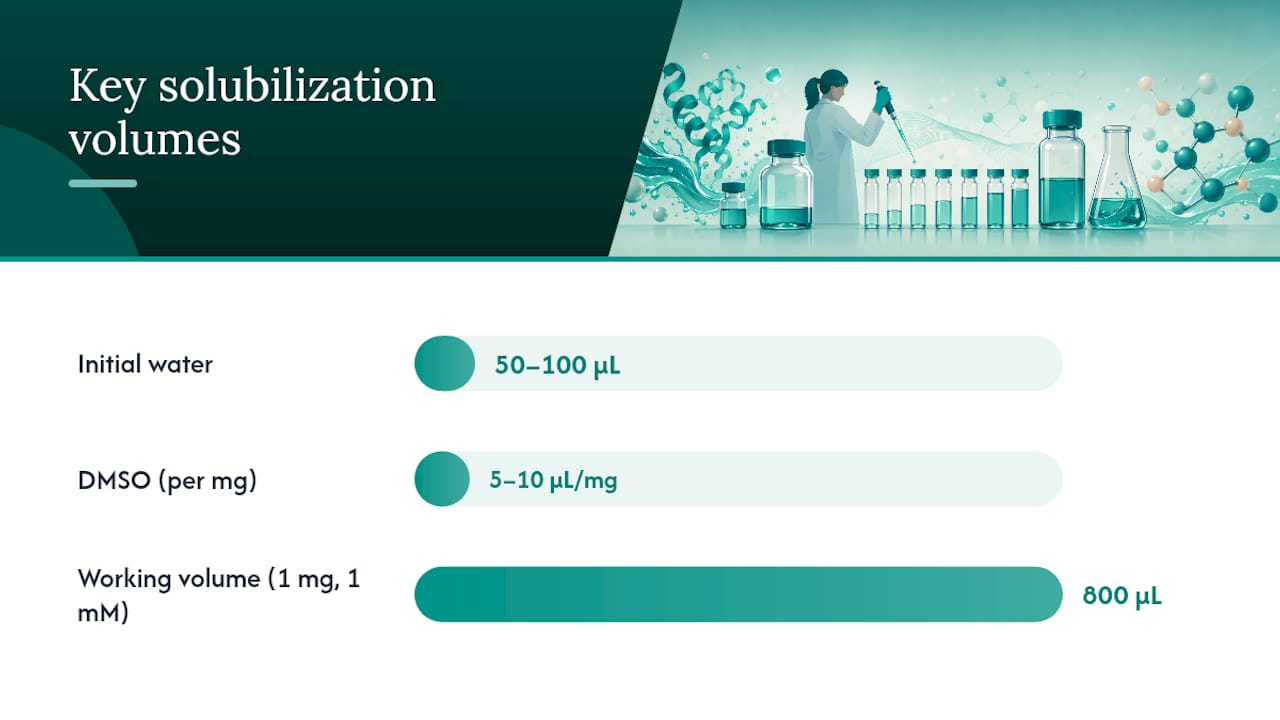
- Add the minimum practical volume of solvent. Start with 50–100 µL of sterile water or BAC water. Cap and roll gently between palms for 30–60 seconds.
- Observe. A clear solution at this stage means water is sufficient. Proceed to concentration calculation and dilution. Persistent cloudiness or a gel-like texture means escalate.
- pH shift. For peptides with pI < 7 (net negative charge at neutral pH), add dilute acetic acid (~0.1 M). For peptides with pI > 7, try dilute ammonium hydroxide (~0.1 M) or phosphate buffer (pH 7.4–8.0).
- Organic pre-wet for hydrophobic sequences. Dissolve the peptide fully in the smallest possible volume of DMSO (typically 5–10 µL per mg). Once dissolved, add aqueous buffer dropwise while gently stirring to avoid local high-concentration precipitation. Document the residual DMSO percentage in the final working solution.
- Sonication as last resort. Place the tube in a bath sonicator for 10–30 seconds. Inspect. Repeat once if needed. Avoid probe sonication directly on the sample.
- Filter if required. Pass through a 0.22 µm sterile syringe filter using aseptic technique (alcohol-swab the filter hub). Use a low-protein-binding membrane (PVDF or PES) to minimize adsorption losses.
Worked concentration example. You have 1 mg of a peptide with MW = 1,250 g/mol and want a 1 mM stock:
- Moles = 1 mg ÷ 1,250 mg/mmol = 0.0008 mmol = 0.8 µmol
- Volume for 1 mM = 0.8 µmol ÷ 1 µmol/mL = 0.8 mL (800 µL)
The benzyl alcohol acts as a preservative and extends the usable window of refrigerated stocks. Always confirm compatibility with your specific assay format before use.
2. How sequence properties predict whether a peptide will dissolve
Sequence composition and net charge at working pH are the strongest predictors of peptide solubility. Knowing these values before opening the vial lets you choose a rational starting solvent rather than cycling through options.
Key molecular determinants:
- Net charge at working pH. Count charged residues: Arg, Lys, His (positive); Asp, Glu (negative); free termini contribute ±1 each. A net charge of ±3 or greater usually predicts good aqueous solubility.
- Isoelectric point (pI). At pI, net charge is zero and solubility is typically lowest. Dissolving at a pH at least 2 units away from pI improves solubility. Free online tools (ExPASy ProtParam, Peptide2.0) calculate pI from sequence in seconds.
- Hydrophobic residue fraction. Sequences where more than 50% of residues are Phe, Leu, Ile, Val, Trp, or Met tend to require organic pre-wetting. A 12-residue peptide with four Phe and three Leu residues falls squarely in this category.
- Length. Longer peptides (>30 residues) accumulate hydrophobic patches and β-sheet propensity, compounding aggregation risk even when individual residues are polar.
- β-sheet and aggregation propensity. Alternating hydrophobic/hydrophilic patterns and polyglutamine or polyalanine stretches signal aggregation risk. Computational tools (TANGO, AGGRESCAN) flag these motifs.
- Modifications. N-terminal acetylation removes the positive charge of the free amine, lowering solubility for already-neutral peptides. C-terminal amidation removes the negative charge of the free carboxyl. Non-natural amino acids (e.g., β-amino acids, D-residues) alter backbone geometry and may require computational screening to predict solubility reliably.
A 20-residue Lys-rich peptide with pI ~10 dissolves readily in water at neutral pH because it carries a strong net positive charge. The 12-residue Phe/Leu-heavy peptide described above will likely need a DMSO pre-wet followed by dilution into phosphate-buffered saline.
Pro Tip: Calculate pI before ordering. If the pI falls between 5 and 8 and the sequence is hydrophobic, request the vendor's ADS to confirm whether an alternate salt form or formulation is available. Counterion identity (e.g., TFA salt) affects intrinsic solubility and is listed on the COA.
3. Which solvents to use, and what your assay can tolerate
Sigma-Aldrich's solubility guidelines recommend water or BAC water as the default starting point, then dilute acetic acid or polar aprotic solvents for hydrophobic sequences, with an explicit warning to verify assay tolerance for any organic solvent before use. The table below maps solvents to use cases and compatibility notes.
| Solvent | Use case | Assay compatibility note |
|---|---|---|
| Sterile / BAC water | Default; basic or charged peptides | Compatible with most assays; BAC water adds preservative |
| Dilute acetic acid (~0.1 M, pH ~3.0) | Acidic or neutral peptides needing low pH | Compatible with most biochemical assays; confirm pH tolerance |
| DMSO | Hydrophobic pre-wet | Keep residual DMSO below assay tolerance; document final % |
| DMF / acetonitrile | Hydrophobic sequences; DMSO-incompatible assays | Higher toxicity; dilute thoroughly; verify cell-assay tolerance |
| TFE / HFIP | Very intractable hydrophobic or amyloid-type peptides | Induce α-helical conformation; avoid for CD/NMR unless controlled |
| Guanidine HCl / urea | Stubborn aggregates | Interfere with enzyme assays; require dialysis or dilution before use |
Assay compatibility rules:
- Cell-based assays. DMSO above 0.1–0.5% (v/v) can affect cell viability and membrane integrity. Determine your assay's specific tolerance empirically and document it.
- Fluorinated alcohols (TFE, HFIP). These dissolve otherwise intractable sequences but bias secondary structure toward α-helix. Use only when solvent effects are acceptable or when a careful solvent-exchange step follows before structural assays.
- Chaotropes. Guanidine HCl and urea disrupt enzyme active sites and interfere with many colorimetric and fluorescence assays. Plan dialysis or lyophilization before downstream use.
Dropwise dilution technique. When transitioning from an organic pre-wet to aqueous buffer, add the aqueous phase dropwise to the organic peptide solution while gently stirring. Reversing the addition order risks local precipitation at the interface.
Pro Tip: When using acetic acid water for reconstitution, use it only when the COA or ADS specifies it. Acetic acid water (~0.6%, pH ~3.0) is appropriate for peptides that require acidic pH to remain in solution, but it carries stability and injection-comfort trade-offs that make it unsuitable as a default solvent.
4. Special handling for short peptides, oxidation-sensitive sequences, and highly hydrophobic peptides
Short peptides (fewer than 8 residues)
Aqueous solubility is often adequate for short peptides, but adsorption to glass and standard polypropylene surfaces becomes a meaningful loss mechanism at low concentrations. Use low-bind microcentrifuge tubes (e.g., Eppendorf LoBind) and low-bind pipette tips.
Oxidation-sensitive residues: Cys, Met, Trp
Cysteine oxidizes to disulfide or sulfenic acid; methionine oxidizes to sulfoxide; tryptophan degrades under UV and oxidative conditions. For these sequences:
- Use degassed, oxygen-free buffers (sparge with nitrogen or argon for 15–20 minutes before use).
- Work on ice or at 4 °C and minimize exposure time.
- Add reducing agents (DTT at 1–5 mM, or TCEP at 0.5–1 mM) only when compatible with downstream assays. TCEP is preferred because it does not interfere with most UV absorbance readings.
- Avoid DMSO for Cys- and Met-containing peptides: DMSO can oxidize these residues directly under certain conditions.
Highly hydrophobic peptides and membrane anchors
Pre-wetting with a minimal volume of DMSO or HFIP before aqueous dilution is the most reliable first intervention for hydrophobic peptides. When fluorinated solvents are used, document the residual solvent concentration and account for potential α-helical bias in any structural readout. For membrane-anchoring sequences, detergent micelles (e.g., SDS, DPC, or LMPG at sub-CMC concentrations) or lipid-mimic environments may be necessary to maintain native conformation during assay.
5. How to run a small-scale solubility test and escalate systematically
Testing on a micro-scale before committing the full vial preserves material and generates reproducible documentation. The escalation ladder below follows the logic of trying the mildest, most easily removed intervention first.
- Weigh out or estimate 1–5% of the vial contents into a low-bind tube. Record mass and lot number.
- Add 50 µL sterile water. Roll gently for 60 seconds. Observe against a dark background under good lighting. Clear = dissolved. Cloudy or gel-like = proceed.
- pH shift. Add 10–20 µL of 0.1 M acetic acid (for acidic/neutral peptides) or 0.1 M NH₄OH (for basic peptides). Roll and observe for 2–3 minutes.
- Organic pre-wet. Add 5–10 µL DMSO to the dry fraction. Dissolve fully, then dilute dropwise with buffer. Check residual DMSO percentage.
- Sonication. Bath-sonicate for 10–30 seconds. Inspect. Repeat once. Stop if no improvement.
- Chaotrope addition. Add guanidine HCl (1–4 M) or urea (4–8 M) to the test fraction. Note: plan a dialysis or dilution step before downstream use.
- Fluorinated alcohol. Add TFE or HFIP in minimal volume. Dissolve, then exchange solvent by lyophilization or dialysis before structural assays. Flag conformational artifact risk.
- Consult vendor or consider re-synthesis. If all steps fail, contact the supplier with full documentation (see Section 10).
Troubleshooting flags:
- Gel formation. Indicates β-sheet aggregation. Try chaotrope or fluorinated alcohol; consider re-synthesis with solubility-enhancing tags.
- Persistent cloudiness after all steps. May indicate particle suspension rather than true dissolution. Filter through 0.22 µm and re-measure absorbance to quantify loss.
- Apparent dissolution but loss of activity. Run an integrity check (HPLC, mass spec) to confirm the peptide is intact and not degraded by the solvent conditions.
Pro Tip: Photograph the test tube against a dark background at each escalation step and timestamp the images. This record is invaluable when contacting the vendor and when reproducing a successful condition at larger scale.
6. Calculating reconstitution volumes and preparing working stocks accurately
The core formula is straightforward. Given mass (mg), molecular weight (g/mol), and desired molar concentration:
Volume (mL) = mass (mg) ÷ [MW (g/mol) × desired concentration (mM)]
Or equivalently: convert mass to micromoles first, then divide by desired concentration in µM to get volume in mL.
Worked example. 1 mg peptide, MW = 2,500 g/mol, target stock = 500 µM:
- Micromoles = (1 mg ÷ 2,500 mg/mmol) × 1,000 = 0.4 µmol
- Volume = 0.4 µmol ÷ 0.5 µmol/mL = 0.8 mL (800 µL)
Reconstitution volume reference (1 mg sample):
Practical stock concentration guidance:
- Set stock concentration high enough that the final assay dilution keeps residual organic solvent below tolerance (e.g., a 10 mM DMSO stock diluted 1:1,000 into assay buffer yields 0.1% DMSO).
- Express stock concentration in µM or mM, not mg/mL, to avoid MW-dependent errors when comparing across peptides.
- Verify stock concentration by UV absorbance (A280 for Trp/Tyr-containing peptides) or by a BCA/Bradford assay where applicable.
7. Storage, sterility, and documentation after reconstitution
Proper post-reconstitution handling is where reproducibility is either secured or lost. Peptide degradation during storage is frequently traced to freeze-thaw cycling, inadequate labeling, or solvent incompatibility with the storage container.
Immediate storage rules:
- Aliquot into single-use volumes immediately after reconstitution. Freeze at ≤ −20 °C for short-term use; use −80 °C for long-term storage or for oxidation-sensitive sequences.
- Avoid freeze-thaw cycles. Each cycle risks aggregation, oxidation, and concentration drift.
- Label every vial: peptide name, lot number, solvent, concentration, % organic, pH, date of reconstitution, and researcher initials.
Sterility and preservatives:
- BAC water (0.9% benzyl alcohol) supports multi-dose storage by inhibiting microbial growth. Refrigerated stocks in BAC water are typically usable for several weeks, though this window depends on peptide stability. Sterility practices for multi-dose vials require consistent aseptic technique at every access.
- Sterile water (no preservative) is appropriate for single-use aliquots or when benzyl alcohol is incompatible with the assay.
- Filter-sterilize through a 0.22 µm low-bind membrane before freezing when the reconstituted solution will be used in cell-based assays.
Record-keeping minimum:
- COA lot number and purity value
- Solvent(s) used, volumes, and order of addition
- Final % organic in working stock
- pH of final solution
- Any solvent-exchange steps performed
- Storage temperature and date
Pro Tip: Record solvent and % organic directly in the methods section of every experiment, not just the lab notebook. Reviewers and collaborators need this information to reproduce your results, and it is the single most commonly omitted detail in peptide reconstitution methods.
8. Validated computational prediction with CamSol-PTM
Sequence-based predictors like CamSol-PTM can reliably screen intrinsic solubility across large peptide design spaces, including sequences containing certain noncanonical amino acids, before any material is synthesized or consumed. The method reports Pearson correlations of approximately 0.6–0.72 between predicted and experimentally measured relative solubility in its validation sets, depending on the dataset.
What CamSol-PTM predicts and its limits:
- Intrinsic solubility based on sequence composition and physicochemical properties, not formulation or counterion effects.
- Extended coverage of noncanonical amino acids enables in silico screening of modified peptide libraries, reducing the experimental validation burden by orders of magnitude in large design spaces.
- Predictions reflect intrinsic sequence properties. They do not account for salt form, counterion, excipients, or the specific buffer conditions of your assay.
Practical integration into the lab workflow:
- Run CamSol-PTM scores on candidate sequences before committing to synthesis. Deprioritize sequences with low predicted solubility unless the application specifically requires them.
- Use predictor scores to rank candidates for small-scale solubility assays, then validate experimentally on 1–5% of the synthesized material before scaling.
- Pair computational predictions with the bench escalation ladder (Section 5) rather than treating a high score as a guarantee of dissolution under your specific conditions.
Pro Tip: Validate computational predictions experimentally on a small fraction of each new batch, even for sequences that scored well in silico. Batch-to-batch variation in counterion, purity, and residual solvent from synthesis can shift real-world solubility away from the predicted value.
9. What to record and what to tell your vendor when a peptide fails to dissolve
A failed dissolution is only useful data if it is fully documented. The checklist below covers what to record before contacting the vendor and what information they need to provide useful guidance.
Documentation checklist:
- Lot number and COA (purity, MW, counterion/salt form)
- Full peptide sequence and any modifications
- Storage history: temperature, duration, number of freeze-thaw cycles, container type
- Exact solvents tried: identity, volume, order of addition, contact time, and temperature
- Observations at each step: clear, cloudy, gel, precipitate, color change
- Analytical evidence if available: HPLC trace, mass spectrum, CD spectrum
What to ask the vendor:
- Request the ADS or lot-specific solubility notes if not already supplied with the COA.
- Ask whether the peptide was supplied as a TFA salt or an alternate counterion (acetate, HCl). TFA salts can reduce aqueous solubility for certain sequences; counterion exchange may help.
- Ask whether alternate salt forms, formulations, or solubility-enhancing modifications (PEGylation, addition of charged tags) are available for re-synthesis.
- Confirm whether the vendor has encountered solubility issues with this sequence in prior lots.
Pro Tip: Include a time-stamped photograph of the vial against a dark background at each dissolution step. Keep a small preserved undissolved fraction in a sealed, labeled tube if possible. Vendors can sometimes run analytical checks on returned material that identify whether the issue is purity, counterion, or sequence-level aggregation. Sequence integrity problems are occasionally the root cause of persistent dissolution failure.
What consistent lab practice actually prevents most dissolution failures
Nearly every persistent reconstitution failure encountered in practice traces back to one of two omissions: skipping the COA/ADS review before opening the vial, or attempting dissolution without knowing the peptide's pI and hydrophobic residue fraction. Both are avoidable in under five minutes.
The habit that prevents most failures is simple: before touching the vial, calculate pI, count hydrophobic residues, and read the vendor's solubility notes. Documenting solvent identity, % organic, and pH in every methods section is not administrative overhead. It is the difference between a result that can be reproduced and one that cannot.
One underappreciated point: researchers often attribute dissolution failure to peptide quality when the actual variable is solvent choice or mechanical handling. Vigorous shaking of a hydrophobic peptide in water will produce aggregates regardless of purity. The protocol matters as much as the material.
Peptasticlabs supports reproducible reconstitution with documented peptides
Fewer solubility surprises start with knowing exactly what is in the vial. Each COA includes lot-specific data that directly informs solvent selection: counterion identity, purity grade, and MW confirmation — the variables that most commonly explain unexpected dissolution behavior.
Researchers working across metabolic, cognitive, tissue repair, longevity, immunology, and cosmetic applications can request a COA before ordering to confirm that the batch documentation meets the requirements of their protocol. For critical experiments, reviewing the COA prior to purchase eliminates a common source of experimental variance. Browse the documented peptide catalog or visit Peptasticlabs to request batch documentation and confirm availability for your research application.
Sources
- Solubility Guidelines for Peptides
- Synthetic Peptide Handling & Storage Protocol
- Peptide solubility - Bachem
- Acetic acid water for peptide reconstitution | PeptideMag