
LL-37 is the sole human cathelicidin antimicrobial peptide, a 37-amino-acid cationic host defense peptide derived from the hCAP18 precursor encoded by the CAMP gene. Its N-terminal sequence begins LLGDFFRKSKEKIGKEFKRIVQRIKDFLRNLVPRTES, and at physiological pH it carries a positive net charge. Researchers and clinicians now prefer the term "host defense peptide" to reflect its dual identity: LL-37 kills bacteria, fungi, and enveloped viruses directly, yet at sub-bactericidal concentrations it modulates cytokine production, recruits immune cells, and accelerates wound re-epithelialization. Processing by serine proteases such as Proteinase 3 (PR3) and kallikrein 5/7 releases the mature peptide from hCAP18 at sites of infection and inflammation.
Core functional categories at a glance:
- Direct antimicrobial: active against Gram-positive and Gram-negative bacteria, selected fungi, and enveloped viruses
- Anti-biofilm: penetrates and disrupts established biofilms; synergy with conventional antibiotics reported
- Immunomodulation: LPS neutralization, chemokine induction (IL-8, MCP-1), macrophage gene-expression reprogramming
- Wound healing: promotes angiogenesis and re-epithelialization in cell and animal models
- Clinical interest: dysregulation linked to rosacea, psoriasis, chronic wounds, and context-dependent oncology findings
- Sourcing note: reproducible results require HPLC-verified ≥99% purity with batch-specific Certificate of Analysis (CoA); Peptasticlabs supplies research-grade LL-37 meeting these standards
Key Takeaways
LL-37 is the sole human cathelicidin, a 37-amino-acid cationic host defense peptide with direct antimicrobial, anti-biofilm, and immunomodulatory functions that operate through distinct concentration-dependent mechanisms.
| Point | Details |
|---|---|
| Definition and origin | LL-37 is a peptide cleaved from hCAP18 (encoded by CAMP) by serine proteases PR3 and KLK5/7. |
| Dual functional roles | At micromolar concentrations LL-37 disrupts bacterial membranes; at lower concentrations it modulates cytokines and recruits immune cells. |
| Key experimental caution | Assay salt concentration and peptide purity critically affect measured activity; always include physiological NaCl controls and verify ≥99% HPLC purity. |
| Primary disease associations | Dysregulation is best documented in rosacea and psoriasis; oncology associations are real but context-dependent and should not be overgeneralized. |
| Peptasticlabs sourcing | Peptasticlabs provides HPLC-verified LL-37 at ≥99% purity with batch CoA and LC-MS confirmation, meeting the QC standards required for reproducible research. |
Table of Contents
- Key biochemical properties of LL-37 at a glance
- How LL-37 is synthesized, processed, and expressed
- How LL-37's structure drives its mechanism of action
- What microbes does LL-37 affect, and how strong is the evidence?
- LL-37's immunomodulatory and wound-healing roles
- Human diseases linked to LL-37 dysregulation
- Therapeutic potential and the main development challenges
- Practical assay design for measuring LL-37 activity
- Standards for research-grade LL-37: purity, QC, and handling
- Open questions and priority research directions
- LL-37 research demands rigor, not just enthusiasm
- Peptasticlabs supports your LL-37 research with verified, documented peptides
- Sources
Key biochemical properties of LL-37 at a glance
| Property | Value / Description |
|---|---|
| Full sequence | LLGDFFRKSKEKIGKEFKRIVQRIKDFLRNLVPRTES |
| Residue count | 37 amino acids |
| Precursor protein | hCAP18 (18 kDa) |
| Encoding gene | CAMP |
| Common synonyms | CAP-18 C-terminal fragment, human cathelicidin |
| Net charge (pH 7) | +5 |
| Secondary structure | Amphipathic α-helix in membrane-mimetic environments |
| Molecular weight | 18 kDa |
Physicochemical properties relevant to bench work:
- Adopts a stable amphipathic α-helix in lipid bilayer-mimetic conditions (e.g., 50% trifluoroethanol, SDS micelles); largely disordered in aqueous buffer alone
- Cationic charge drives electrostatic attraction to negatively charged bacterial membranes and LPS
- Hydrophobic moment sufficient for membrane insertion and permeabilization
- Soluble in water and dilute acetic acid; use low-binding tubes at concentrations near the MIC to minimize adsorption losses
Natural fragments and sequence variants: Proteolytic processing does not always yield full-length LL-37. Shorter fragments including RK-31, KS-30, and KR-20 are generated in skin and other tissues by kallikrein-family proteases. Each fragment carries a distinct potency and cytotoxicity profile. RK-31 retains substantial antimicrobial activity with reduced hemolytic potential; KR-20 is less active against bacteria but retains some immunomodulatory function. When designing experiments or interpreting published MIC data, confirm which fragment or analog was used, because activity differences between fragments can be substantial.
How LL-37 is synthesized, processed, and expressed
Understanding the biosynthetic pathway clarifies why LL-37 concentration, fragment identity, and local tissue context vary so widely across experimental and clinical settings.
Stepwise processing pathway:
- CAMP transcription: The CAMP gene on chromosome 3p21.3 is transcribed in neutrophils, epithelial cells, and other cell types; transcription is strongly induced by 1,25-dihydroxyvitamin D3 (calcitriol) acting through vitamin D response elements in the CAMP promoter, as well as by NF-κB-activating inflammatory signals and short-chain fatty acids from commensal bacteria.
- hCAP18 translation and storage: The 18 kDa prepropeptide hCAP18 is synthesized and stored in secondary (specific) granules of neutrophils; epithelial cells secrete it constitutively at lower levels.
- Secretion: Neutrophil degranulation at infection sites or epithelial secretion releases hCAP18 into the extracellular space.
- Proteolytic cleavage: Serine proteases, primarily Proteinase 3 (PR3) in neutrophils and kallikrein 5/7 (KLK5/7) in skin, cleave the cathelin domain from the C-terminal region to release mature LL-37. Kallikrein-mediated processing in the stratum corneum also generates the shorter fragments RK-31 and KS-30.
- Local concentration dynamics: Mature LL-37 accumulates at micromolar concentrations in neutrophil extracellular traps (NETs) and at wound surfaces; systemic plasma concentrations are far lower and largely below bactericidal thresholds.
Tissue and cell expression map:
- Neutrophils: highest constitutive expression; primary source during acute infection
- Epithelial cells (skin, airway, gut): inducible expression; key barrier defense role
- Mast cells and NK cells: express LL-37 and can release it in response to pathogen signals
- Monocytes/macrophages: lower baseline expression, upregulated by vitamin D and infection
Vitamin D receptor (VDR) signaling is the best-characterized transcriptional regulator of CAMP expression. Ionic strength and pH at the site of secretion affect helical induction and therefore the peptide's local activity, independent of concentration. Serine protease activity itself is regulated by local pH and protease inhibitors, adding another layer of post-translational control over the final active fragment profile.
How LL-37's structure drives its mechanism of action
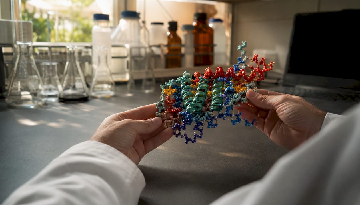
LL-37's amphipathic α-helix and cationic charge enable both direct membrane permeabilization and high-affinity binding to negatively charged bacterial components such as LPS. This structural duality is the foundation of its multifaceted host defense roles.
Membrane disruption models
Three mechanistic models describe how cationic amphipathic peptides permeabilize bacterial membranes, and LL-37 shows features consistent with more than one:
- Carpet model: peptide molecules accumulate on the membrane surface in a detergent-like manner until a threshold coverage causes membrane dissolution; favored at high peptide-to-lipid ratios
- Toroidal pore model: peptide helices insert into the bilayer and curve the lipid monolayers inward, forming transient water-filled pores lined by both lipid headgroups and peptide
- Detergent-like/micellar solubilization: at very high concentrations, membrane fragments are solubilized into mixed peptide-lipid micelles
Against Gram-negative bacteria, LL-37 must first cross the outer membrane by displacing divalent cations from LPS; this step is electrostatically driven. Gram-positive bacteria lack an outer membrane, so LL-37 acts directly on the cytoplasmic membrane, though the thick peptidoglycan layer can slow access. The net result is rapid membrane depolarization, leakage of cytoplasmic contents, and cell death.
Receptor-mediated and signaling actions
At sub-bactericidal concentrations, LL-37 functions through defined receptor interactions rather than direct membrane lysis:
- FPR2 (formyl peptide receptor 2): mediates chemotaxis of neutrophils and monocytes; also triggers anti-inflammatory lipoxin-like signaling at low peptide concentrations
- P2X7 receptor: LL-37 activates P2X7 on macrophages, triggering IL-1β processing and inflammasome activation
- EGFR transactivation: LL-37 can transactivate epidermal growth factor receptor in epithelial cells, contributing to wound re-epithelialization
- MrgX2 (Mas-related GPCR): expressed on mast cells; mediates mast cell degranulation in response to LL-37, relevant to allergic and inflammatory contexts
Concentration dependence and ionic strength effects
The functional outcome of LL-37 exposure is strongly concentration-dependent. At nanomolar to low micromolar concentrations, receptor-mediated immunomodulatory and chemotactic effects dominate. At higher micromolar concentrations, direct membrane permeabilization and cytotoxicity emerge. Physiological ionic strength (150 mM NaCl) reduces helical induction and lowers measured bactericidal activity relative to low-salt assay conditions, a critical caveat when extrapolating in vitro MIC data to in vivo settings. Many published bactericidal concentrations were measured in low-salt media and likely overestimate killing efficiency at mucosal surfaces or in serum.
What microbes does LL-37 affect, and how strong is the evidence?
LL-37 is broadly active against many Gram-positive and Gram-negative bacteria, certain fungi, and some enveloped and non-enveloped viruses; it also disrupts biofilms and reduces biofilm viability, with anti-biofilm and antimicrobial findings varying substantially with assay conditions.
Representative activity data from published literature (interpret with assay-condition caveats):
| Organism / Target | Activity Type | Key Caveat |
|---|---|---|
| E. coli, P. aeruginosa | Bactericidal (MIC reported in low-salt media) | Activity reduced at physiological NaCl; strain variation documented |
| S. aureus, S. epidermidis | Bactericidal; anti-biofilm | MIC values vary widely by strain and medium; MRSA strains may show reduced susceptibility |
| Candida albicans | Antifungal (membrane disruption) | Concentrations required often higher than for bacteria |
| Enveloped viruses (HSV, influenza) | Membrane disruption of viral envelope | Non-enveloped viruses generally less susceptible |
| P. aeruginosa biofilm | Biofilm dispersal and viability reduction | Synergy with tobramycin reported in some studies; concentration and timing matter |
| LPS (endotoxin) | Neutralization / binding | Reduces macrophage TNF-α stimulation; protective in mouse endotoxemia models |
Anti-biofilm activity in detail: LL-37 penetrates biofilm matrices and reduces viable cell counts within established biofilms. Dispersal activity has been documented for P. aeruginosa and S. aureus biofilms in vitro. Synergy with conventional antibiotics, including tobramycin and colistin, has been reported in multiple experimental systems, though the magnitude of synergy is assay- and strain-dependent. Researchers should treat published biofilm MIC/MBEC (minimum biofilm eradication concentration) values as condition-specific benchmarks rather than universal thresholds.
Strain-to-strain variability is substantial. Some clinical isolates of P. aeruginosa and S. aureus have developed resistance mechanisms including modification of membrane charge (via MprF or dltABCD operons) that reduce LL-37 binding. Reporting exact strain identifiers, passage history, and assay conditions is therefore non-negotiable for reproducible biofilm work.
LL-37's immunomodulatory and wound-healing roles
LL-37 is a pleiotropic host defense peptide that recruits immune cells, modulates cytokine production in both pro- and anti-inflammatory directions, promotes angiogenesis, and accelerates re-epithelialization in wound models. These non-microbicidal roles are arguably as important as direct killing for understanding its biology, and they operate at concentrations well below the bactericidal range.
Specific immunomodulatory actions with documented evidence:
- Chemokine induction: LL-37 induces IL-8 (CXCL8) and MCP-1 (CCL2) in epithelial cells and monocytes, driving neutrophil and monocyte recruitment to infection sites
- Chemokine receptor upregulation: increases surface expression of CXCR4 and CCR2 on immune cells, amplifying chemotactic responses
- LPS and LTA neutralization: binds and neutralizes bacterial endotoxins, reducing macrophage TNF-α stimulation and protecting mice against lethal endotoxemia in experimental models
- NET stabilization: associates with DNA in neutrophil extracellular traps, stabilizing NETs and potentially amplifying innate immune signaling; this same complex formation underlies autoimmune pathology in psoriasis
- P2X7 activation: triggers inflammasome-dependent IL-1β release in macrophages
- Anti-inflammatory modulation: at low concentrations via FPR2, LL-37 can suppress excessive inflammatory responses, illustrating its concentration-dependent bidirectional immunomodulatory profile
Macrophage gene-expression profiling confirms that LL-37 reprograms macrophages toward a phenotype with altered cytokine output, enhanced phagocytosis, and modified toll-like receptor responsiveness. Mouse models of lethal endotoxemia show that LL-37 administration reduces mortality, providing in vivo support for the LPS-neutralization mechanism. These protective effects are robust across multiple experimental systems.
Wound-healing evidence is strongest in cell culture and rodent models. LL-37 activates EGFR in keratinocytes, accelerating migration and proliferation. It also stimulates angiogenesis through VEGF-A induction, which supports granulation tissue formation. The peptides in immune modulation literature provides useful complementary context on how host defense peptides interact with immune signaling networks more broadly.

Human diseases linked to LL-37 dysregulation
LL-37 dysregulation is implicated in dermatologic diseases (rosacea, psoriasis), chronic wounds and infections, respiratory mucosal inflammation, and has context-dependent links to cancer biology. The strength of evidence varies considerably across these categories.
Skin and dermatology
Rosacea and psoriasis represent the best-supported associations. In rosacea, abnormal cathelicidin processing by kallikrein 5 generates atypical LL-37 fragments that trigger exaggerated innate immune responses, driving the characteristic facial erythema and inflammatory papules. Elevated total LL-37 is also documented at rosacea lesion sites. In psoriasis, LL-37 forms complexes with self-DNA released from damaged keratinocytes; these complexes activate plasmacytoid dendritic cells via TLR7/9, initiating the type I interferon cascade that sustains the psoriatic inflammatory loop. This mechanism positions LL-37 as a direct molecular link between barrier damage and autoimmune amplification.
Mucosal inflammation and chronic infections
At mucosal surfaces, LL-37 is upregulated in chronic nasal inflammatory disease and other inflamed mucosa. Deficient LL-37 production in the airway is associated with increased susceptibility to bacterial colonization, particularly in cystic fibrosis, where high-salt airway surface liquid inactivates the peptide. Chronic wounds show both elevated LL-37 (from persistent neutrophil infiltration) and impaired processing that may generate less active fragments, contributing to biofilm persistence.
Oncology context
The oncology picture is genuinely complex and should not be oversimplified. In some cancer models (ovarian, lung), LL-37 has been reported to promote tumor cell proliferation and angiogenesis, while in others it shows cytotoxic or anti-proliferative effects. The direction of effect depends on tumor type, local concentration, receptor expression profile, and the immune microenvironment. Clinical dysregulation data confirm context-dependent effects in cancer, and researchers should treat any single-model finding with appropriate caution.
Microbiome implications
LL-37 shapes microbial community composition at mucosal surfaces by selectively suppressing susceptible species while sparing more resistant commensals. Disruption of this selective pressure, through either excess or deficient LL-37, can shift microbiome balance in ways that may perpetuate inflammatory disease. This area remains an active and undercharacterized research front.
Therapeutic potential and the main development challenges
LL-37 is conceptually attractive as a resistance-mitigating host defense peptide but requires engineering to be clinically viable. Its multi-target mechanism makes resistance evolution harder for bacteria than with single-target antibiotics, and its immunomodulatory properties offer adjunct therapeutic value beyond direct killing.
Delivery modalities compared by suitability and risk:
- Topical (wound/skin): highest feasibility; local concentration control is achievable; systemic toxicity risk is low; proteolytic degradation by wound proteases remains a challenge; formulation in hydrogels or nanoparticles can extend half-life
- Inhaled (pulmonary): promising for cystic fibrosis and ventilator-associated pneumonia; local delivery bypasses systemic exposure; salt concentration in airway surface liquid must be managed; nebulization may fragment the peptide
- Systemic (IV/subcutaneous): highest risk profile; rapid proteolytic turnover in plasma limits half-life; concentration-dependent cytotoxicity to host cells at therapeutic doses is a real constraint; systemic off-target immunostimulation is poorly characterized
Primary development challenges:
- Proteolytic instability: LL-37 is rapidly degraded by serine and metalloproteinases in plasma, wound fluid, and mucosal secretions; half-life in serum is short
- Concentration-dependent cytotoxicity: the therapeutic window between bactericidal and hemolytic/cytotoxic concentrations is narrow for the native peptide
- Off-target immunostimulation: mast cell activation via MrgX2 and inflammasome activation via P2X7 can produce unintended inflammatory effects at non-target sites
- Manufacturing and purity requirements: peptide synthesis at scale requires rigorous QC; impurities and oxidation products can introduce cytotoxicity artifacts
- U.S. regulatory pathway: peptide therapeutics require IND filing with the FDA; demonstrating safety, purity, and potency for a cationic AMP with immunomodulatory activity involves extensive preclinical toxicology
Synthetic analog strategies address several of these challenges simultaneously. D-amino acid substitutions confer protease resistance without altering the overall helical geometry. Lipidation improves membrane affinity and can lower the effective concentration needed for bactericidal activity. Stapled or constrained helices maintain secondary structure in aqueous environments, reducing the dependence on membrane-mimetic conditions for activity. Each modification carries trade-offs: D-amino acid substitution may alter receptor-mediated signaling; lipidation increases hemolytic potential; stapling adds synthetic complexity and cost. Researchers evaluating analogs should report which modifications were made and test both antimicrobial and cytotoxic endpoints in parallel.
Practical assay design for measuring LL-37 activity
Use standardized MIC and time-kill protocols adapted for cationic peptides, and include salt and serum controls to mimic physiological ionic strength. This single design choice separates interpretable data from artifacts.
Assay selection and design
MIC determination: Broth microdilution (CLSI M07 framework) is standard, but cationic peptides require modifications. Use Mueller-Hinton broth supplemented to physiological NaCl (150 mM) as a parallel condition alongside standard low-salt broth to quantify the salt sensitivity of your specific peptide batch. Agar-based MIC methods are generally less suitable for LL-37 because peptide adsorption to agar can substantially reduce free peptide concentration.
Time-kill assays: Confirm bactericidal kinetics at 1×, 2×, and 4× MIC. Rapid killing (within 1–2 hours) is characteristic of membrane-active peptides and distinguishes LL-37 from bacteriostatic agents.
Biofilm assays: Use both biomass endpoints (crystal violet staining) and viability endpoints (CFU enumeration or resazurin reduction) because LL-37 can reduce viability without fully dispersing the biofilm matrix. Report peptide concentration, contact time, growth phase of the biofilm, and medium composition.
Assay checklist for reproducible LL-37 experiments:
- Positive control: a well-characterized AMP (e.g., melittin or polymyxin B at known MIC)
- Negative control: scrambled-sequence peptide at matched concentration to control for non-sequence-specific effects
- Hemolysis assay: test against human red blood cells at the same concentration range used in antimicrobial assays; report HC50 (50% hemolytic concentration) relative to MIC
- HPLC/LC-MS purity confirmation: verify peptide identity and purity from the same batch used in experiments; degradation products can produce false-positive or false-negative activity
- Endotoxin testing (LAL assay): required for any cell-based assay to exclude LPS contamination as a confounding variable
- Adsorption control: measure free peptide concentration in your assay vessel after equilibration but before adding bacteria, using low-binding polypropylene tubes
Data-interpretation caveats
Peptide adsorption to polystyrene plates and medium components (serum proteins, divalent cations) reduces free peptide concentration in ways that are difficult to quantify without direct measurement. Always report the exact peptide form used: free acid vs. C-terminal amidation, acetylation status, and counterion (TFA vs. acetate), because these affect charge, solubility, and activity. TFA counterions at high concentrations can themselves be cytotoxic in cell assays; acetate-form peptides are preferred for cell-based work.
Standards for research-grade LL-37: purity, QC, and handling
For reproducible results, use HPLC-verified peptides at ≥99% purity with a batch-specific Certificate of Analysis (CoA) and supporting LC-MS identity verification. This is not a preference; it is a reproducibility requirement. Impurities and degradation products in lower-grade preparations introduce cytotoxicity artifacts that can invalidate both antimicrobial and cell-based assay results.
QC checklist for researchers ordering LL-37:
- CoA with HPLC chromatogram showing ≥99% purity by peak area
- LC-MS confirmation of molecular weight matching the expected sequence (monoisotopic or average mass, clearly stated)
- Counterion specification (TFA or acetate); request acetate form for cell-based assays
- Lyophilized storage at -20°C or -80°C; avoid moisture exposure
- Recommended reconstitution solvent (typically sterile water or 10–20% acetonitrile in water for initial dissolution, then dilution into buffer)
- Stability data or expiry information for the lyophilized and reconstituted forms
Handling notes:
- Prepare single-use aliquots immediately after reconstitution; avoid repeated freeze-thaw cycles, which promote aggregation and oxidation of methionine residues
- Use low-binding polypropylene tubes for all dilutions at or near MIC concentrations
- Account for adsorption losses when working below 10 µg/mL; consider measuring actual free peptide concentration by HPLC or BCA assay after equilibration
Pro Tip: Request the impurity profile alongside the HPLC chromatogram, not just the purity percentage. A preparation reporting 99% purity with a single large impurity peak at a cytotoxic mass is more problematic than one with several trace peaks well below 0.5%. The impurity identity matters as much as the total purity figure.
Vendors that provide stability data, impurity profiles, and counterion specifications alongside standard CoA documentation give researchers the information needed to design clean experiments. Peptasticlabs documents these parameters for its cataloged peptides, consistent with the research-grade peptide standards that minimize batch-to-batch variability in published work.
Open questions and priority research directions
The highest-priority gaps in LL-37 research, where new work would have immediate translational impact:
- Accurate local tissue concentration mapping: plasma and bulk tissue measurements do not reflect the micromolar concentrations at infection foci or wound surfaces where LL-37 acts. Imaging mass spectrometry and spatially resolved proteomics could provide the first reliable maps of local LL-37 distribution in human tissue.
- Standardized in vivo biofilm models: current animal models for biofilm-associated infection vary widely in implant material, bacterial species, and inoculum; a consensus model would allow direct comparison of LL-37 and analog efficacy across laboratories.
- Receptor-specific signaling dissection: FPR2, P2X7, EGFR, and MrgX2 each mediate distinct downstream effects, but most published studies use whole-cell systems where multiple receptors are active simultaneously. Receptor knockout or selective antagonist studies in relevant primary cell types would clarify which receptor drives which outcome at which concentration.
- Long-term safety of repeated topical and inhaled exposure: single-dose toxicology data exist, but chronic exposure studies in relevant animal models are sparse. This gap is the primary bottleneck for advancing inhaled LL-37 analogs toward IND-enabling studies.
- Microbiome impact quantification: co-culture models combining human epithelial cells with defined microbial communities would allow controlled measurement of how LL-37 concentration shifts microbiome composition, a question with direct relevance to inflammatory bowel disease and atopic dermatitis research.
Translational experiments with the highest near-term feasibility include dose-toxicity window studies in murine wound infection models using topically applied stabilized analogs, and combinatorial antibiotic-LL-37 studies in biofilm reactor systems with clinically relevant strains. Both are achievable with current methodology and would directly inform formulation decisions.
LL-37 research demands rigor, not just enthusiasm
The field has accumulated a substantial body of mechanistic and clinical association data on LL-37, and the therapeutic rationale is genuinely strong. A peptide that kills bacteria through physical membrane disruption, neutralizes endotoxin, recruits immune cells, and promotes tissue repair addresses multiple failure modes of conventional antibiotics simultaneously. That combination is rare.
The problem is that enthusiasm for this profile has sometimes outpaced methodological rigor. Published MIC values for LL-37 span orders of magnitude across studies using nominally the same organism, largely because assay salt concentration, peptide purity, and adsorption controls are inconsistently reported. Immunomodulatory findings from transformed cell lines are regularly extrapolated to in vivo predictions without validation in primary cells or animal models. And the oncology literature contains contradictory findings that are difficult to reconcile because the experimental systems are too heterogeneous to compare.
The reproducibility problem is solvable. It requires transparent reporting of peptide source, batch purity, exact sequence and modification state, assay conditions, and negative controls in every publication. Researchers who treat these as optional details rather than core methods are the primary source of noise in this field. The science of LL-37 is strong enough to stand on its own when the experiments are done carefully. It does not need to be oversold.
Peptasticlabs supports your LL-37 research with verified, documented peptides
Reproducible LL-37 research starts with a peptide you can trust. Peptasticlabs supplies research-grade LL-37 verified to ≥99% purity by HPLC, with batch-specific Certificates of Analysis and LC-MS identity confirmation available on request. Every lot is independently tested, and counterion specifications are documented so you can select acetate-form material for cell-based assays without ambiguity.

The vendor checklist researchers should apply before ordering any LL-37 preparation: HPLC chromatogram with ≥99% purity by peak area, LC-MS molecular weight confirmation, counterion specification, lyophilized storage conditions, and recommended reconstitution protocol. Peptasticlabs provides all of these as standard documentation, not as add-ons. For researchers who need bulk quantities or custom batch documentation for institutional compliance, wholesale options are available. Browse the research-grade peptide catalog to review available compounds, purity documentation, and ordering options, or visit Peptasticlabs for full QC and CoA details.
Sources
The references below represent the highest-value starting points for LL-37 research, covering mechanism, clinical associations, experimental methodology, and translational challenges.
- The Potential of Human Peptide LL-37 as an Antimicrobial and Anti-Biofilm Agent
- Human antimicrobial/host defense peptide LL-37 may prevent the spread of a local infection through multiple mechanisms: an update | Inflammation Research | Springer Nature Link
- The human antimicrobial peptide LL-37 is a multifunctional modulator of innate immune responses
- LL-37, the master antimicrobial peptide, its multifaceted ...
- LL-37 - Wikipedia
This article is general information, not a substitute for advice from a qualified doctor. Consult a qualified healthcare professional about your own circumstances before acting on anything here.