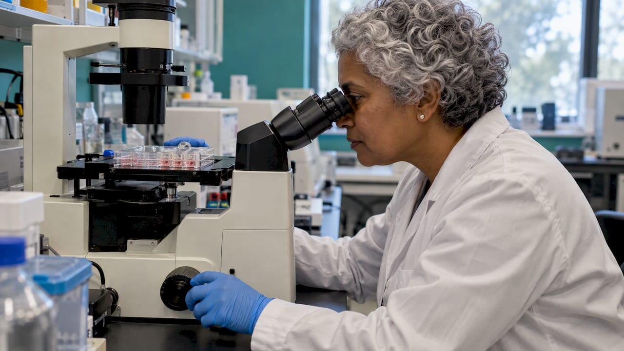
A wound healing assay, also called a scratch assay, measures how fast a cell monolayer closes a defined cell-free gap. The gap closure is read as a surrogate for collective cell migration. The method is simple and low cost, and its results are only as reliable as the standardization behind them: seeding density, scratch width, serum conditions, imaging coverage and a way to separate migration from proliferation. This guide covers each of those points for researchers setting up or troubleshooting the assay.
TL;DR
- The assay creates a gap in a confluent monolayer, images it over time and quantifies closure. It reports planar collective cell migration, not the full in vivo repair process.
- Reproducibility depends on seeding density, scratch width and imaging coverage. Imaging at least 76% of the scratched area gives more robust data than a single field of view.
- Proliferation confounds closure. Serum starvation, mitotic inhibitors such as mitomycin C or inserts help isolate migration, and each needs validation in the chosen cell line.
- Pipette-tip scratches are cheap and fast. Inserts, wound makers and robotic platforms trade cost for lower variability.
- Closure is calculated as (a - b) / a x 100, where a is initial width and b is width after closure.
Table of Contents
- What a wound healing assay measures
- Wound creation methods compared
- Step-by-step protocol outline
- Controls and conditions that decide reliability
- Separating migration from proliferation
- Imaging and quantification
- Common failure points
- Choosing test articles for the assay
- Perspective
- Research materials for migration studies
- FAQ
- Sources
What a wound healing assay measures
The in vitro wound-scratch assay is a widely used model for studying collective cell migration, a process that contributes to wound closure and re-epithelialisation. A defined gap is generated in a confluent monolayer. Closure over time is monitored as a surrogate readout of repair.
The assay is popular for practical reasons. It is simple, low cost and adaptable, which has made it a foundational tool for early-stage wound-healing research and therapeutic screening. A 2026 narrative review examined 199 published studies and identified 73 relevant to wound healing.
It provides quantitative data on cell motility, which makes it suited to evaluating drugs, growth factors or genetic modifications. According to the Nature Protocols method paper, it is particularly suitable for studying cell-matrix and cell-cell interactions in migration, and it is compatible with live-cell imaging.

What it does not measure
Gap closure reflects planar collective migration. It does not recapitulate the integrated inflammatory, vascular, metabolic and extracellular matrix-dependent processes that govern repair in vivo. Compounds acting through antioxidant, anti-inflammatory, angiogenic or matrix-modulating pathways can be underestimated or misclassified by migration-only readouts. Design the study with that boundary stated up front, and pair the scratch assay with complementary endpoints where the mechanism requires it.
Wound creation methods compared
The wide array of techniques is a documented cause of poor reproducibility between laboratories. A 2026 review describes a technical hierarchy of three categories.
| Method | Examples | Strength | Limitation |
|---|---|---|---|
| Mechanical | Pipette tip, cell scraper | Accessible, cheap, fast | Operator-dependent variability |
| Semi-automated | Culture inserts, wound maker devices | Improved reproducibility | Added cost, insert handling |
| Fully automated | Robotic platforms | High precision, high throughput | Equipment access |
A Frontiers methods review notes that the culture insert increases reproducibility between replicates, while the pipette-tip scratch is much cheaper and faster.
The creation method can change biology as well as variance. In a fibroblast comparison, migration was significantly reduced when wounds were made with a cell exclusion method. Cortisol added to the medium reduced migration only in scratched monolayers, not in exclusion-created wounds. A scratch damages cells and matrix at the edge. An exclusion gap does not. Report the method in every figure legend and do not compare absolute closure rates across methods.
Step-by-step protocol outline
The basic sequence is to scratch a monolayer, image at the start and at regular intervals, and compare images to quantify the migration rate. The parameters below follow a standardized approach published in 2023.
- Seed cells. In the published standardized method, 60,000 keratinocytes (HEKa) or 10,000 primary dermal fibroblasts per well were placed in a 24-well plate and grown to 90% confluence, usually within 3 days.
- Synchronize. Replace growth medium with basal medium lacking growth factors, FBS or supplements for 18 to 24 h, holding cells at the quiescence stage (G0).
- Scratch. Introduce a full-thickness scratch with a 200 µL or 1000 µL pipette tip, using a consistent angle and pressure.
- Wash. Rinse to remove detached cells and debris, then add the test medium.
- Image at time zero. Record the initial gap before any closure.
- Image at intervals. Use fixed positions and fixed time points. Live-cell imaging allows continuous tracking.
- Quantify. Measure gap width or area at each time point and calculate closure.
Controls and conditions that decide reliability
Seeding density and confluence
Create a confluent monolayer of 90 to 95% confluence and keep the seeding number constant for each cell line. The required number depends on cell type and dish size.
The 2023 standardization study specifies seeding at 31,578 cells/cm² for HEKa and 5,263 cells/cm² for primary human fibroblasts. Density also affects scratch geometry: in primary dermal fibroblasts, higher densities produced a uniform sheet with less control over initial scratch width.
Tip size
Small 200 µL tips create narrower scratches and a faster closure rate. That makes it harder to detect variables with a medium impact on closure. If the aim is to detect modest effects, a wider scratch gives a longer window.
Serum conditions
Serum concentration is a variable to be set per cell type, not a default. In one fibroblast study, the best-performing serum level differed by cell origin:
| Fibroblast type | Serum condition with best migration |
|---|---|
| Pulmonary | 1% and 0.5% FCS |
| Juvenile skin | Significant increase only at 2% FCS |
| Adult dermal | 4% FCS |
| Aortic adventitial (AoAF) | Higher FCS (4% and 10%) |
Pulmonary and juvenile skin fibroblasts migrated better than AoAF and adult dermal fibroblasts without added FCS. Adding ascorbic acid to different FCS concentrations reduced migration capability. Run a serum titration for any new cell line before testing compounds.
Control wells to include
- Untreated (medium only) control for baseline closure.
- Vehicle control matching the solvent of the test article.
- Positive control with a known migration-promoting stimulus, where one is established for the cell line.
- Proliferation control, such as a mitotic inhibitor arm (see below).
- Technical replicates in several wells, with several independent experiments.
Separating migration from proliferation
The key limitation of the assay is possible confoundment by other cellular phenotypes, which can cause misinterpretation of outcomes. Cells dividing at the edge of the gap close it as effectively as cells moving into it.
Common mitigation steps are serum starvation, mitotic inhibitors and commercial inserts, which help isolate migratory behavior from proliferative effects. One published approach scores influences on both migration and cell death while normalizing cell growth with mitomycin C (MMC) pre-treatment.
MMC is not neutral in every model. In that study, MMC pre-treatment significantly affected the closure rate in KKU-055 (cholangiocarcinoma) and A549 (lung) cells beyond blocking proliferation, implying it can perturb migration itself. Cell line choice is therefore a critical step in any MMC protocol.
Practical consequences:
- Validate the inhibitor dose and exposure time in each cell line.
- Compare closure with and without the inhibitor.
- Keep assay duration short relative to the doubling time.
- Add a viability readout, so cell death is not read as reduced migration.
Imaging and quantification
Imaging coverage
A single field of view gives highly variable scratch area data. In the 2023 standardization study, five sequential digital images at 4x magnification covered on average 76 ± 2.6% of the scratched length in each well of a 24-well plate. Total scratch area per replicate produced much more robust data than the average area per field. The study's recommendation is to document at least 76% of the scratched area.
Calculating closure
The percentage of wound closure is (a - b) / a x 100, where a is the width of the wound in images taken after scratching and b is the width following closure.
Worked example: initial width 600 µm, width at 24 h 240 µm. Closure = (600 - 240) / 600 x 100 = 60%.
Where possible, use area rather than width, and normalize each well to its own time-zero value, so scratches of different initial size remain comparable.
Reporting checklist
- Cell line, passage range and seeding density.
- Serum or medium composition during the assay.
- Wound creation method, tool size and any inserts.
- Fraction of the scratch imaged and number of fields.
- Time points and the formula used.
- Proliferation control and its result.
- Number of independent experiments.
Lack of standardization makes results hard to compare. Jonkman et al. recommend general guidelines for sample preparation, microscope requirements, image acquisition and image analysis of gap size and closure rate.
Common failure points
The assay is amenable to variables that produce false-positive and false-negative data. Variability lowers sensitivity, and removing its sources improves detection of variables with wide-ranging effects.
| Problem | Likely cause | Corrective step |
|---|---|---|
| Uneven scratch width | Manual pressure, angle or tip drift | Use a guide or ruler, one operator, or an insert |
| Cells detach or peel | Weakly adherent cell type | Reconsider the assay for the model, or use coated plates |
| Closure too fast to compare | Narrow scratch, high proliferation | Wider tip, lower serum, mitotic inhibitor |
| High well-to-well variance | Single field imaged | Image at least 76% of the scratch |
| Subjective analysis | Manual tracing | Fixed analysis rules or automated software |
Manual wound creation introduces variability, and the assay is not ideal for non-adherent or weakly adherent cell types.
Choosing test articles for the assay

The assay is used to screen peptides, growth factors and small molecules for effects on migration. Test-article quality affects the data: identity, purity and batch traceability determine whether a response can be attributed to the intended compound. Documentation such as an HPLC purity report and a Certificate of Analysis supports that attribution. For related method planning, see the guides on selecting peptides for cell culture and in vitro peptide testing methods. Assays for antimicrobial activity are covered in the article on antimicrobial peptide assays.
Perspective
In our experience of supplying research materials, assay variability is usually discussed in terms of cells, tips and imaging. The test article is the fourth variable, and it is the one a laboratory can fix at purchase.
Our position is that a scratch assay result is interpretable only when the compound is characterized. That is why each batch we supply is independently tested by HPLC and mass spec, verified to ≥99% purity, with traceable batch documentation and Certificates of Analysis available on request.
Our Tissue & Repair and Cosmetic Science lines are organized for the kinds of repair and skin-model studies in which migration assays are common. We designed the documentation so a researcher can record the batch used alongside the cell line, density and scratch method.
Tintastic
Research materials for migration studies
Peptastic Labs supplies independently tested, research-grade peptides across eight product lines: Metabolic, Cognitive & Neuro, Tissue & Repair, Longevity, Cosmetic Science, Blends, Hormone & Reproductive, and Ancillaries & Reagents. Each is HPLC-verified to ≥99% purity, with batch documentation and Certificates of Analysis on request. Bulk and wholesale options are available.
- Browse the full peptide catalogue for Tissue & Repair and Cosmetic Science compounds.
- Request the Certificate of Analysis for the batch before recording it in your methods.

Orders are placed online, including pre-filled Telegram or WhatsApp checkout. Products are supplied for laboratory research.
FAQ
What is the difference between a wound healing assay and a scratch assay?
The terms describe the same in vitro method. A gap is made in a confluent monolayer and closure is imaged over time. "Wound healing assay" is the broader label, and it also covers non-scratch methods such as exclusion inserts.
How long should a scratch assay run?
It depends on the cell line's migration speed and doubling time. Image at time zero and then at regular intervals until the gap is substantially closed in controls. Shorter runs limit the contribution of proliferation. Set the time points in a pilot experiment.
Can a scratch assay show that a compound heals wounds?
No. It shows an effect on planar collective migration in a monolayer. It does not capture inflammatory, vascular or matrix-dependent processes, so it is best used for early-stage screening and followed by models that include those processes.
Is a pipette tip or an insert better?
Tip scratches are cheaper and faster but operator-dependent. Inserts raise reproducibility between replicates at higher cost. Choose by the effect size you need to detect and the number of replicates you can run.
How many images are needed per well?
One field is not enough. In a 24-well plate, five sequential 4x images covered about 76% of the scratch length on average, and the study recommends documenting at least 76% of the scratched area.
Sources
- The In Vitro Wound-Scratch Assay: Applications, Technical Considerations (PubMed, 2026)
- In vitro scratch assay: a convenient and inexpensive method (Nature Protocols, 2007)
- A Robust and Standardized Approach to Quantify Wound Closure (PMC, 2023)
- Wound healing assay protocol (Abcam)
- Novel Analytical Platform for Robust Identification of Cell Migration (Scientific Reports, 2020)
- In vitro Cell Migration, Invasion, and Adhesion Assays (Frontiers, 2019)
- Comparison of in vitro scratch wound assay experimental methods (PMC, 2023)
- Primary Human Fibroblasts in Culture Switch to a Myofibroblast-Like Phenotype (PMC, 2019)
- An introduction to the wound healing assay using live-cell microscopy (PMC, 2014)