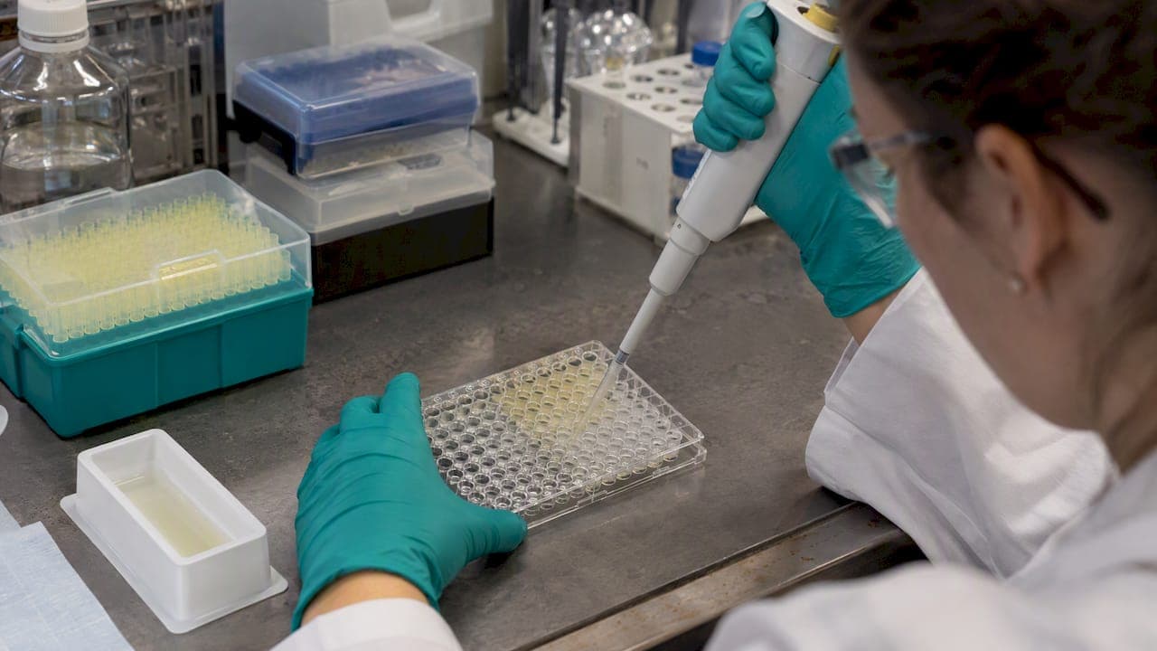
Antimicrobial peptide (AMP) assays fall into three groups: activity assays (MIC and related growth tests), safety assays (hemolysis and cytotoxicity) and mechanism assays (membrane disruption studies). A defensible AMP paper or internal screen needs all three. This guide covers the standard setup for each, the handling errors that most often distort results, and the documentation an Australian lab should have in place before ordering peptides.
TL;DR
- Run the MIC by broth microdilution, and record inoculum, medium, incubation time and temperature with every result.
- Use polypropylene plates for cationic peptides. Polystyrene can bind them and shift concentrations.
- Pair every activity assay with a hemolysis or cell viability assay. Activity alone says little about a candidate.
- Prepare peptide stocks fresh. Verify identity and purity (HPLC, mass spec) before interpreting any result.
- In Australia, research peptides are for in vitro laboratory use only. Human-use supply is regulated by the TGA.
Table of Contents
- What an antimicrobial peptide assay measures
- The MIC assay by broth microdilution
- Modifications for cationic peptides
- Handling and storage
- Safety assays: hemolysis and cytotoxicity
- Going past MIC: kinetics and mechanism
- From discovery to validation
- Common errors that distort AMP results
- Research-use rules in Australia
- Our perspective
- Sourcing peptides for assay work
- FAQ
- Sources
What an antimicrobial peptide assay measures
An AMP is a short amino acid sequence made by organisms across the tree of life. Most act on the microbial membrane. Reported mechanisms fall into pore-forming models (barrel-stave, toroidal) and non-pore-forming models (carpet, detergent-like). In each, peptides interact with membrane lipids and the cell leaks its contents and lyses.
Because the mechanism is physical, assay design matters more than it does for many small molecules. Charge, surface binding, medium salts and plastic type all change what the peptide does in the well. A review of tools for evaluating new AMPs makes the same point: the result depends heavily on method.
Three questions structure the work:
| Question | Typical assay | Output |
|---|---|---|
| Does it inhibit or kill the organism? | Broth microdilution MIC, MBC, time-kill, growth curves | MIC (lowest concentration with no visible growth), kill kinetics |
| Does it spare host cells? | Hemolysis, mammalian cell viability | HC50, cell viability, safety margin |
| How does it act? | Membrane permeabilization, microscopy, cryo-electron tomography | Mechanism evidence |
The MIC assay by broth microdilution
The MIC is the entry point. Under standardized protocols, broth microdilution identifies the lowest antimicrobial concentration that inhibits visible bacterial growth after 16 to 24 hours of incubation at 37°C. The usual starting inoculum is about 5 × 10^5 CFU/mL in cation-adjusted Mueller-Hinton broth.
Step-by-step outline
- Confirm peptide identity and purity from the Certificate of Analysis (CoA) for the batch you are using.
- Prepare the peptide stock fresh on the day of the assay.
- Make serial twofold dilutions in the test medium.
- Adjust the bacterial culture to the target inoculum and add it to each well.
- Include growth control (no peptide), sterility control (no bacteria) and a reference antibiotic control.
- Incubate 16 to 24 hours at 37°C.
- Read the lowest concentration with no visible growth. Repeat in independent experiments.
Replicates matter. MIC values move in twofold steps, so a single well difference is within normal variation. Report the number of independent runs and how you handled disagreements, such as taking the mode.
Why MIC values are not interchangeable
MIC is inherently variable. Inoculum size, incubation time and medium composition, including cation concentration, all influence it. Values generated under CLSI and EUCAST conditions are not directly comparable. If you compare your numbers with a published paper, check the method section first. The same logic appears in Adelaide University's mycology methods page, which lists inoculum, incubation time and temperature, and medium type among the factors that shape antifungal susceptibility results.
A record sheet should capture, for every plate:
- Organism, strain ID and growth phase
- Inoculum density (CFU/mL, confirmed by plate count)
- Medium and lot, including cation adjustment
- Plate material and supplier
- Incubation time and temperature
- Peptide batch number and CoA reference
Modifications for cationic peptides
Most AMPs are cationic, and that creates a practical problem. Cationic peptides bind to polystyrene, which makes results unreliable in standard 96-well plates. The Hancock Lab method, a modification of classical microtiter broth dilution, uses polypropylene for that reason. Polypropylene plates and tubes reduce peptide loss and give more reproducible concentration data.

The same lab's method page is a good reference for how to prepare dilutions of cationic peptides. A comparison of broth microdilution methods for cationic peptides tested 90 clinical isolates against six peptides and is useful when deciding between method variants.
Keep modifications justified
Standards bodies are cautious on this point. CLSI guidance on modifying susceptibility testing methods discourages changes to reference broth microdilution. Where a peptide needs an adjustment, the change must be scientifically justified. It must never be used to lower MIC values and favor one compound over another. In practice:
- Change one variable at a time and report it.
- Run the reference method in parallel where possible.
- State the reason for the change in the methods section.
Handling and storage
Peptide handling is where many irreproducible results start. The standard approach, described in a study of Australian scorpion peptides, is to prepare fresh stocks on the day of the assay. If storage is unavoidable, keep peptide at -80°C in the dark. Repeated freeze-thaw cycles change concentration and activity.
Checklist for reconstitution and storage:
- Reconstitute only what the assay needs.
- Aliquot single-use volumes if you must store.
- Use low-binding polypropylene tubes.
- Label each aliquot with batch number and date.
- Discard thawed leftovers rather than refreezing.
Safety assays: hemolysis and cytotoxicity
Antimicrobial activity is only one aspect of peptide evaluation. Hemolytic and cytotoxic assays are essential safety checkpoints. A promising candidate shows a wide margin: effective antimicrobial concentrations sit well below those that cause hemolysis or reduce human cell viability. Hemolysis is commonly summarized as HC50, the concentration causing 50% hemolysis.
A simple way to express the margin is a ratio of HC50 to MIC. A larger ratio indicates better selectivity. Report the red blood cell source, the cell concentration, the incubation time and the positive control (typically a detergent giving 100% lysis) so the ratio can be reproduced.
| Parameter | What to record |
|---|---|
| Cell type | Species and source of erythrocytes or the cell line used |
| Cell density | Final concentration in the assay |
| Exposure | Time and temperature |
| Controls | Buffer-only (0% lysis), detergent (100% lysis) |
| Readout | HC50 and the concentration range tested |
For cell viability, use a standard readout appropriate to your lab, and test over the same concentration range as the MIC series so the two datasets line up.
Going past MIC: kinetics and mechanism
The MIC has limits. It can miss regrowth, and it does not separate bacteriostatic from bactericidal action. Researchers note that growth curve analysis and cryo-electron tomography are increasingly used to visualize membrane disruption and confirm mechanism at the cellular level.
Common follow-up assays:
- Minimum bactericidal concentration (MBC): plate wells showing no growth to see whether cells were killed or only inhibited.
- Time-kill or growth curves: sample over time at multiples of the MIC to show kill rate and regrowth.
- Membrane permeabilization assays: dye uptake or leakage readouts that test the membrane-disruption hypothesis.
- Imaging: electron microscopy or cryo-electron tomography for direct visualization.
Biofilm and wound-relevant conditions are another direction. A 2026 paper on a synthetic peptide targeting MDR wound pathogens notes that biofilms, high ionic strength and protease-rich exudates limit conventional antimicrobials, so assays in simple broth can overestimate real-world performance. Test in conditions that reflect your question.
From discovery to validation
Computational tools help prioritize sequences, but they do not replace bench work. A review of AMP discovery to application states that after candidates are identified, experimental validation is essential. The next step is typically synthesis by solid-phase peptide synthesis (SPPS), then the assays above.

Synthesis quality feeds directly into assay quality. A peptide with truncated sequences or unknown salts gives unreliable MICs, because the real concentration of active peptide is unknown. Identity by mass spectrometry and purity by HPLC are the baseline. High-resolution mass spectrometry is routine in AMP papers, for example in a study on gramicidin S derivatives against Gram-negative ESKAPE pathogens, where HRMS is described alongside HPLC. For more on reading purity data, see our guide to peptide purity standards in research and our overview of in vitro peptide testing methods.
Common errors that distort AMP results
| Error | Effect | Fix |
|---|---|---|
| Polystyrene plates for cationic peptides | Peptide loss, inflated MIC | Polypropylene plates and tubes |
| Old or repeatedly thawed stocks | Altered concentration and activity | Fresh stocks, or -80°C single-use aliquots |
| Unrecorded inoculum or medium | Results cannot be compared | Log every parameter per plate |
| Method changes without justification | Artificially favorable MICs | Justify, report and run the reference method |
| MIC without safety data | Misleading candidate ranking | Add hemolysis and viability assays |
| Unverified peptide identity | Unknown active content | Batch CoA with HPLC and mass spec data |
Research-use rules in Australia
In Australia, peptides sold for research are treated as non-therapeutic laboratory reagents. The Therapeutic Goods Administration (TGA) prohibits supply for human use unless the product is an approved medicine on the Australian Register of Therapeutic Goods (ARTG). Research-grade material must be documented as for in vitro laboratory use only. A separate explainer makes the related point that a peptide not on the ARTG but marketed for human use is an unapproved therapeutic good.
For an AMP lab this means:
- Keep purchase records, intended-use statements and CoAs on file.
- Do not use research material in humans or animals outside an approved ethics and regulatory pathway.
- Check import paperwork before ordering. See our notes on documents Australian labs must attach to import research peptides and HS codes for peptides.
This is general information, not legal advice. For a specific import or institutional question, speak with your institution's compliance or biosafety office.
Our perspective
We supply research peptides, so we see how often an assay problem is a material problem. A twofold MIC shift can come from the plate, the medium or the stock. It can also come from a peptide whose identity or purity was never confirmed. We cannot fix your protocol, but we can remove one variable.
Our approach is documentation first. Each batch is independently tested by HPLC and mass spectrometry, with traceable batch records. Compounds in the catalog are verified to ≥99% purity, and a Certificate of Analysis is available on request. Tie that CoA to your lab notebook entry for every assay run.
Our practical advice for AMP work:
- Request the CoA before the first experiment, not after an anomalous result.
- Record the batch number next to each MIC and hemolysis dataset.
- Keep assay conditions fixed between batches, so a batch change is the only variable.
- Treat research material as in vitro only, in line with TGA rules.
— Tintastic
Sourcing peptides for assay work
Peptastic Labs lists more than 22 independently tested compounds across metabolic, cognitive and neuro, tissue and repair, longevity, cosmetic science, blends, hormone and reproductive, and ancillaries and reagents lines. Each is HPLC-verified to ≥99% purity and supplied with batch documentation. Bulk and wholesale options are available for larger programs. Browse the full catalogue and check availability for your compound of interest, or see the Peptastic Labs homepage for ordering details. For order size planning, read about documents that decide peptide MOQ.
FAQ
What is an antibacterial assay?
It is a laboratory test that measures whether a compound inhibits or kills bacteria. The most common format is broth microdilution, which reports the MIC. Follow-up tests such as MBC and time-kill assays show whether the effect is bactericidal and how fast it acts.
What is the mechanism of action of antimicrobial peptides?
Most AMPs disrupt microbial membranes. Proposed models include pore-forming (barrel-stave, toroidal) and non-pore-forming (carpet, detergent-like) mechanisms. Both lead to leakage of cell contents and lysis.
Why use polypropylene plates for AMP MIC assays?
Cationic peptides bind to polystyrene, so the concentration in solution can fall below the intended value. Polypropylene limits that loss and improves reproducibility.
Is an MIC enough to judge a peptide?
No. MIC does not capture regrowth or separate bacteriostatic from bactericidal action. Add hemolysis or cytotoxicity assays to calculate a safety margin, and use kinetics or imaging to study mechanism.
Can research peptides be used in humans in Australia?
No. Research-grade peptides are for in vitro laboratory use only. The TGA prohibits supply for human use unless a product is approved and on the ARTG, as outlined in this 2026 research guide.
Sources
- New and old tools to evaluate new antimicrobial peptides, PMC
- Modified MIC Method for Cationic Antimicrobial Peptides, Hancock Lab
- In Vitro Susceptibility Tests for Cationic Peptides, PMC
- CLSI: Modification of antimicrobial susceptibility testing methods
- Membrane interactions and biological activity of antimicrobial peptides from Australian scorpion
- Antimicrobial peptide mechanisms, Nature Communications
- Antimicrobial peptides: from discovery to developmental applications, PMC
- Gramicidin S derivatives against ESKAPE pathogens
- Synthetic antimicrobial peptide targeting MDR wound pathogens, Journal of Materials Chemistry B
- Laboratory Methods, Mycology, Adelaide University
- Are Peptides Legal in Australia? 2026 Research Guide
- Are Peptides Legal in Australia? The 2026 TGA Reality