
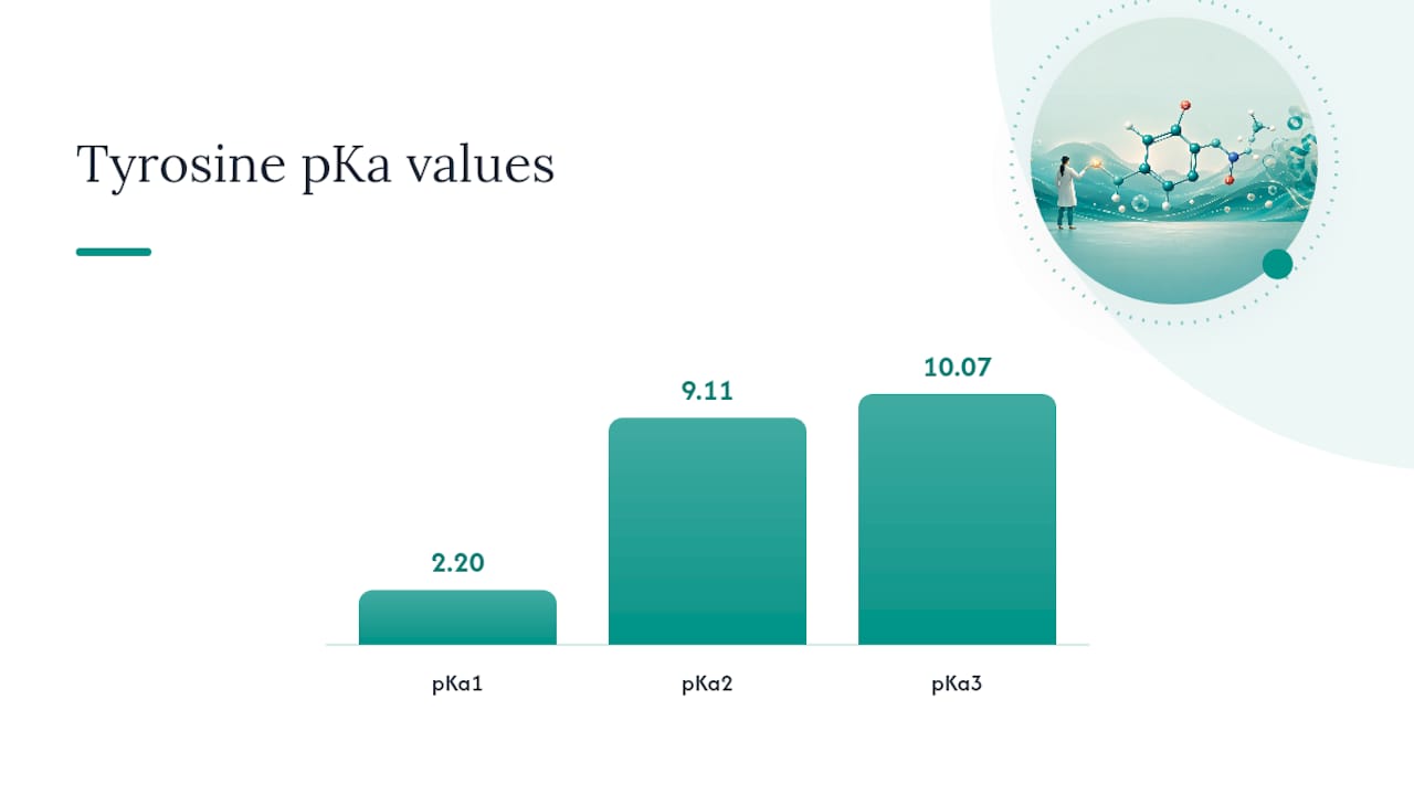
The isoelectric point of L-tyrosine is ≈5.66, calculated by averaging the two pKa values that bracket the net-neutral (zwitterionic) species. The three standard macroscopic pKa values are: carboxyl group ≈ 2.20, amino group ≈ 9.11, and phenolic side chain ≈ 10.07. Because the neutral species sits between the carboxyl deprotonation (pKa1 = 2.20) and the amino deprotonation (pKa2 = 9.11), those two values are averaged: pI = (2.20 + 9.11) / 2 = 5.655 ≈ 5.66. The phenolic pKa of 10.07 does not participate in this average because it flanks a species that already carries a net negative charge.
5.66 — the pI of L-tyrosine, derived from pKa1 (carboxyl, 2.20) and pKa2 (amino, 9.11), with the phenolic side chain pKa (10.07) excluded from the average.
Key Takeaways
The isoelectric point of L-tyrosine is ≈5.66, calculated by averaging pKa1 (2.20) and pKa2 (9.11), the two values flanking the net-neutral zwitterion.
| Point | Details |
|---|---|
| pI of L-tyrosine | ≈5.66, from averaging pKa1 (carboxyl, 2.20) and pKa2 (amino, 9.11). |
| Bracketing rule | Average only the two pKa values that flank the net-neutral species, not all three. |
| Phenolic pKa excluded | The side-chain pKa (10.07) flanks a net-negative species, so it is not included in the average. |
| Use numerical methods for peptides | Set up a Henderson–Hasselbalch charge-balance expression and solve Q(pH) = 0 by bisection. |
| Microenvironment shifts pI | Hydrogen bonding and local dielectric can move the effective phenolic pKa by one or more pH units in proteins. |
Table of Contents
- How do you calculate the isoelectric point of tyrosine step by step?
- Which pKa values should you average for a three-pKa amino acid?
- How does tyrosine's charge state change across pH?
- When does simple averaging fail, and how do you solve pI numerically?
- Why does pI matter biologically, and what shifts it in proteins?
- An editorial note on precision and pragmatics
- Sources
How do you calculate the isoelectric point of tyrosine step by step?
The arithmetic is straightforward once you map each pKa to its ionizable group and identify the neutral species. The values below come from PubChem CID 6057 and are consistent with the worked examples in Pearson's instructional resource.
| Ionizable group | pKa | Net charge contribution at low pH |
|---|---|---|
| Carboxyl (–COOH) | 2.20 | Protonated → 0 |
| Amino (–NH3⁺) | 9.11 | Protonated → +1 |
| Phenolic –OH | 10.07 | Protonated → 0 |
Step-by-step calculation:
- Write the protonation states. At very low pH, all three groups are protonated: the carboxyl is –COOH (neutral), the amino is –NH3⁺ (+1), and the phenol is –OH (neutral). Net charge = +1.
- Deprotonate in pKa order. Raising pH past 2.20 removes the carboxyl proton, giving –COO⁻. Net charge drops to 0 — this is the zwitterion (neutral species).
- Identify the bracketing pKas. The neutral species exists between pKa1 (2.20) and pKa2 (9.11). Those two values bracket it.
- Compute pI. pI = (pKa1 + pKa2) / 2 = (2.20 + 9.11) / 2 = 11.31 / 2 = 5.655, rounded to 5.66.
- Confirm significant figures. For classroom work, two decimal places (5.66) is standard. Three significant figures (5.655) is appropriate when reporting experimental data.
The phenolic pKa (10.07) is excluded because it flanks the singly deprotonated anion, not the neutral species. Averaging it in would shift the result to ≈6.39, which is incorrect.
Which pKa values should you average for a three-pKa amino acid?
The general rule: average the two pKa values that flank the net-neutral species. This applies to any amino acid with three ionizable groups, including tyrosine and cysteine. MasterOrganicChemistry's tutorial walks through this logic clearly for both cases.
To apply the rule, list the full protonation sequence from fully protonated (lowest pH) to fully deprotonated (highest pH), tracking net charge at each step. The neutral species is the one with net charge = 0. The pKa immediately below it and the pKa immediately above it are the two you average.
- Tyrosine: Sequence is +1 → 0 → −1 → −2. Neutral species sits between pKa1 (2.20) and pKa2 (9.11). Average those two → pI ≈ 5.66.
- Cysteine: pKa values are ≈1.96 (carboxyl), ≈8.18 (thiol), ≈10.28 (amino). Sequence is +1 → 0 → −1 → −2. Neutral species sits between pKa1 (1.96) and pKa2 (8.18). Average those two → pI ≈ 5.07.
Pro Tip: The most common student error is averaging the wrong pair — often the two highest pKa values or all three. Always trace the charge sequence first. The neutral species is your anchor; the pKas on either side of it are the only ones that matter.
How does tyrosine's charge state change across pH?
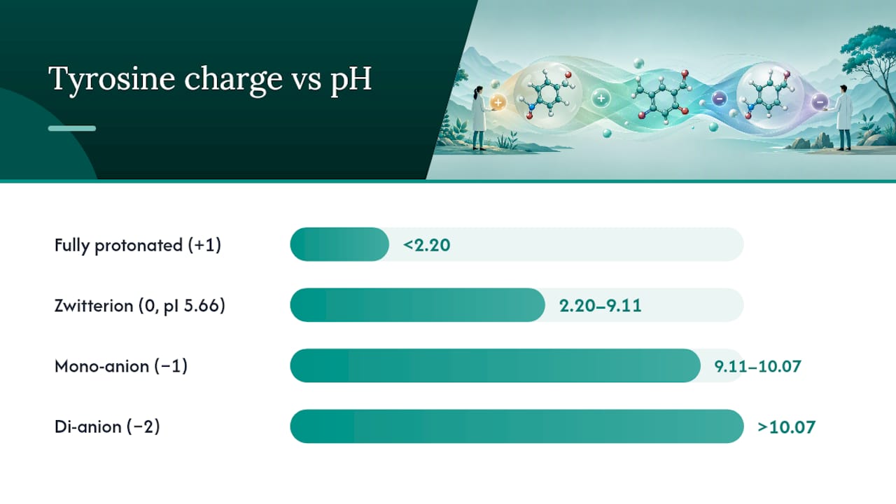
Tyrosine passes through four distinct charge states as pH rises from strongly acidic to strongly basic. The zwitterion — the net-neutral form — is the dominant species near pH 5.66.
| Species | Protonation pattern | Net charge | Approximate pH region |
|---|---|---|---|
| Fully protonated cation | –COOH, –NH3⁺, –OH | +1 | pH < 2.20 |
| Zwitterion (neutral) | –COO⁻, –NH3⁺, –OH | 0 | pH 2.20–9.11 |
| Mono-anion | –COO⁻, –NH2, –OH | −1 | pH 9.11–10.07 |
| Di-anion | –COO⁻, –NH2, –O⁻ | −2 | pH > 10.07 |
The zwitterion spans a wide pH window (roughly 2.20 to 9.11) because the carboxyl and amino pKas are far apart. At exactly pH 5.66, the concentrations of the +1 and −1 species are equal and minimal, so net charge is zero. This is why tyrosine's solubility is lowest near its pI — reduced electrostatic repulsion between molecules allows aggregation.
The pI is not simply the midpoint of the pH scale. It is the pH at which the sum of all positive and negative charge contributions across every ionizable group equals zero — a charge-balance condition, not a geometric average of all pKa values.
When does simple averaging fail, and how do you solve pI numerically?
Arithmetic averaging works well for isolated amino acids with well-separated pKa values. It breaks down in three situations: when ionizable groups are closely spaced in pKa (within ~1–2 units), when multiple groups interact electrostatically, or when you are working with a peptide or protein carrying many ionizable residues.
For those cases, the Henderson–Hasselbalch equation provides the charge fraction for each group at a given pH. The net charge expression for tyrosine is:
Q(pH) = −[1 / (1 + 10^(pKa1 − pH))] + [10^(pKa2 − pH) / (1 + 10^(pKa2 − pH))] + [10^(pKa3 − pH) / (1 + 10^(pKa3 − pH))]
where pKa1 = 2.20, pKa2 = 9.11, pKa3 = 10.07. Setting Q(pH) = 0 and solving for pH gives the precise pI.
Numerical root-finding algorithm:
- Define Q(pH) as the sum of Henderson–Hasselbalch charge fractions for all ionizable groups.
- Set a pH search range (e.g., 0–14).
- Evaluate Q at the midpoint; if Q > 0, shift the lower bound up; if Q < 0, shift the upper bound down.
- Repeat (bisection) until |Q| < 0.001 or the pH interval is smaller than 0.001 units.
- Report the converged pH as pI.
For a three-pKa amino acid like tyrosine, bisection converges in fewer than 50 iterations. For peptides with 10 or more ionizable residues, this approach is the only reliable method.
For research-grade precision — especially when pI governs a separation or a binding experiment — numerical root-finding is not optional. Arithmetic averaging is a classroom shortcut, not a measurement protocol.
The RSC micro-ionization study documents how macro-pKa values themselves are averages over microstates, which introduces additional uncertainty when precision is required. For metal-binding or enzyme active-site work, micro-pKa analysis and spectrophotometric decomposition are the correct approach.
Pro Tip: When building a charge-balance expression for a peptide, include the N-terminus (pKa ≈ 8) and C-terminus (pKa ≈ 3.1) as separate ionizable groups alongside every ionizable side chain. Omitting either terminus shifts the computed pI.
Why does pI matter biologically, and what shifts it in proteins?
The solution-state pI of 5.66 is a starting point, not a fixed property once tyrosine is incorporated into a peptide or protein. Local microenvironment — hydrogen bonding networks, neighboring charged residues, and reduced dielectric constant in hydrophobic pockets — can shift the effective phenolic pKa by one or more pH units, as documented in the ab initio study of L-tyrosine molecular properties. This matters practically in several contexts:
- Electrophoresis and isoelectric focusing: Proteins migrate to their pI in an IEF gel. Using solution-state amino acid pI values to predict protein migration is unreliable; sequence-based computational tools or experimental measurement are required. For peptide-level work, sequence integrity directly affects predicted pI.
- Tyrosine phosphorylation: Phosphorylation of the phenolic –OH eliminates that ionizable group and adds a phosphate (pKa ≈ 1 and ≈6), shifting the local charge state substantially at physiological pH.
- Solubility and formulation: Peptides are least soluble near their pI. Knowing the pI guides buffer selection for reconstitution and storage.
Experimental factors that shift observed pKa and pI values:
- Temperature: pKa values for amino and carboxyl groups shift by roughly 0.01–0.03 units per degree Celsius. Always note the temperature when reporting pKa data.
- Ionic strength: High salt screens electrostatic interactions, compressing apparent pKa differences.
- Binding partners: Metal ions, cofactors, or adjacent residues can stabilize or destabilize the protonated form of the phenol, shifting its effective pKa by 1–3 units.
When your experiment depends on solubility, fractionation, or charge-dependent binding, measure pI directly rather than relying on literature values. In vitro peptide testing methods provide practical frameworks for this kind of characterization. Verifying reagent purity before any pI-sensitive assay is equally important; a Certificate of Analysis confirms identity and purity before you commit to experimental conditions.
An editorial note on precision and pragmatics
Arithmetic averaging is sufficient for homework and quick benchmarks. For any experiment where pI governs a separation, a solubility cutoff, or a binding interaction, that shortcut introduces error that compounds with every additional ionizable group in the molecule. The bracketing rule gives you the right answer for isolated amino acids; numerical root-finding gives you the right answer for everything else. Consult primary data sources — the CRC Handbook of Chemistry and Physics and Lehninger Principles of Biochemistry — when you need verified literature pKa values, and cross-check against PubChem CID 6057 for solution-state macroscopic constants. If the experiment depends on it, measure it.
Sources
- L-Tyrosine | C9H11NO3 | CID 6057 - PubChem
- Ab initio study of molecular properties of l-tyrosine
- Micro-ionization constants for tyrosine - Transactions of the Faraday Society
- Isoelectric Points of Amino Acids (and How To Calculate Them) - MasterOrganicChemistry
- Calculate the isoelectric point of tyrosine (Y) | Pearson