
KPV, the tripeptide Lys-Pro-Val, enters cells through the PepT1 transporter and suppresses NF-κB signaling once inside. The strongest evidence comes from human cell lines and murine DSS/TNBS colitis models, where oral and injected KPV consistently lowers disease-activity scores and pro-inflammatory cytokine expression. No robust human randomized trials exist, and dosing in people remains undefined, so KPV peptide research today sits firmly at the mechanistic and translational stages, not the clinical one.
TL;DR:
- KPV enters cells via PepT1, with expression upregulated during gut inflammation, affecting its absorption and intracellular activity in diseased tissue.
- Its anti-inflammatory effect involves blocking NF-κB p65 nuclear translocation by competitively inhibiting importin-α3 binding, supported by broad concentration ranges in vitro.
- Preclinical evidence shows KPV reduces colitis severity in mouse DSS and TNBS models, working independently of melanocortin receptor pathways, with nanoparticle delivery improving colon targeting.
- Human dosing, safety, and pharmacokinetics remain unestablished, and peptide sourcing must include verification of purity and batch data to ensure reproducibility.
- Future research should prioritize human safety trials, standardized uptake measurements, and innovative stabilization methods to advance clinical translation.
Table of Contents
- KPV Peptide Research: What the Tripeptide Actually Is
- How KPV Enters Cells and Shuts Down NF-κB
- Preclinical Evidence: Colitis Models and Delivery Innovations
- Where KPV Research Is Headed: Therapeutic Areas and Endpoints
- Human Evidence Gaps and Safety Signals to Watch
- Designing Reproducible KPV Experiments: A Practical Checklist
- Sourcing Verified KPV for Laboratory Research
- KPV Peptide Comparisons: How It Stacks Up Against Related Analogs
- Beyond NF-κB: Other Signaling Pathways KPV May Influence
- Measuring KPV Bioavailability: Why It's Harder Than It Sounds
- Toxicity and Side Effects Observed in Preclinical Models
- Next-Generation KPV: Stability and Activity Enhancement Research
- What Would Actually Move KPV Research Forward
- Sourcing Research-Grade KPV for Your Next Study
- Primary Literature and Resources for Continued Reading
- Sources
- FAQ
KPV Peptide Research: What the Tripeptide Actually Is
KPV is the C-terminal tripeptide fragment of alpha-melanocyte-stimulating hormone (α-MSH), consisting of lysine, proline, and valine in that sequence. Researchers isolated it from the parent hormone because it retains anti-inflammatory activity independent of the melanocortin receptors that α-MSH normally engages. That distinction matters for anyone designing a study: KPV works even in melanocortin-1 receptor (MC1R) deficient models, which points to a separate intracellular pathway rather than classic receptor-ligand signaling on the cell surface.
Its small size (three amino acids, molecular weight around 358 daltons) makes it a useful minimal pharmacophore for studying which part of α-MSH drives the anti-inflammatory signal, stripped of the pigmentation and appetite effects tied to full-length melanocortin peptides.
Formulations and routes used in the literature:
- Oral gavage in rodent colitis models, often the free peptide or a nanoparticle-encapsulated version to survive gastric and intestinal proteases.
- Topical application in skin and wound-healing studies, where the peptide is delivered directly to the lesion site.
- Intraperitoneal or intracolonic injection, used in mechanistic studies to bypass oral degradation entirely and confirm local tissue effects.
Handling notes matter more for KPV than for larger peptides. As a short, unprotected sequence, it is vulnerable to peptidase cleavage in gut lumen and serum, which is why researchers studying oral delivery have moved toward protective carriers. Standard peptide storage practices apply: lyophilized aliquots at negative 20°C, minimal freeze-thaw cycling, and reconstitution in sterile buffer immediately before use. Because the peptide is short and highly polar, verifying identity and purity by HPLC and mass spectrometry before an experiment begins is not optional for reproducible work.
How KPV Enters Cells and Shuts Down NF-κB
The uptake step is where KPV's story gets interesting for anyone used to thinking of peptides as either "too big to get in" or "just diffusing passively." KPV instead hitches a ride on PepT1, the proton-coupled di/tripeptide transporter normally responsible for absorbing dietary peptide fragments in the small intestine. This transporter-mediated entry is a large part of why KPV research is a template for peptide pharmacology more broadly.
PepT1 is not uniformly expressed. It shows up strongly in intestinal epithelial cells and, notably, becomes upregulated in inflamed colon tissue during active colitis, which research on PepT1 expression in inflamed gut has documented across multiple studies. That inflammation-dependent expression pattern means KPV's intracellular availability can differ substantially between healthy and diseased tissue, and it is a variable serious study designs need to measure rather than assume.
Uptake kinetics have been characterized in Caco2-BBE cells, a standard intestinal epithelial line. The foundational PepT1 uptake study reports an hPepT1 Km of approximately 160 μM for KPV in this system, a relatively low affinity that still supports meaningful intracellular accumulation at the concentrations used in most in vitro assays.
What happens once KPV gets inside the cell:
- KPV interferes with the interaction between the NF-κB subunit p65 and importin-α3, the nuclear transport protein p65 normally requires entering the nucleus.
- Mechanistic work on this interaction indicates KPV competitively blocks that p65/importin-α3 binding, preventing p65 from translocating into the nucleus.
- Without nuclear p65, transcription of NF-κB target genes, including TNF-α, IL-6, and IL-1β, drops measurably in treated cells.
KPV's effective concentration range in cell culture studies spans a broad range from low nanomolar to moderate micromolar concentrations, according to the PepT1 uptake and NF-κB inhibition study. That four-log spread is unusually wide for a single peptide and reflects differences across cell types, exposure duration, and readout assay, which is exactly why assay standardization matters when comparing results across labs.
For anyone designing an in vitro protocol, this mechanism has a direct implication: a luciferase reporter assay for NF-κB activity, paired with a Western blot for IκBα degradation and p65 nuclear localization, gives you the cleanest read on whether KPV is doing what the literature says it does in your specific cell system.
Preclinical Evidence: Colitis Models and Delivery Innovations
The DSS (dextran sulfate sodium) and TNBS (trinitrobenzene sulfonic acid) murine colitis models form the backbone of KPV's preclinical case. Both chemically induce colonic inflammation that mimics features of human inflammatory bowel disease, and both have produced consistent results when KPV is administered orally or by enema.
The original PepT1-mediated uptake study demonstrated that oral KPV reduced colitis severity in both DSS and TNBS models. A separate murine colitis analysis reported improved body weight recovery, reduced myeloperoxidase (MPO) activity as a marker of neutrophil infiltration, and lower histologic inflammation scores. Disease-activity index (DAI) scores, which combine weight loss, stool consistency, and rectal bleeding into a single composite measure, improved consistently across these studies alongside reduced expression of TNF-α, IL-6, and IL-1β at the mRNA level.
One finding stands out as particularly important for mechanism-focused researchers: KPV retained its anti-inflammatory activity in MC1R-deficient mice. Since α-MSH's classic anti-inflammatory signaling runs through melanocortin receptors, KPV working without a functional MC1R confirms that the PepT1/NF-κB pathway operates independently of the receptor system the parent hormone relies on.
What the preclinical evidence shows, in sequence:
- DSS models show reduced DAI scores and preserved colon length, a standard proxy for inflammation severity, within days of starting KPV treatment.
- TNBS models replicate the anti-inflammatory signal with a different inflammatory trigger, strengthening confidence that the effect is not an artifact of one induction method.
- MC1R-deficient mice confirm receptor-independent action, isolating the PepT1/NF-κB pathway as the operative mechanism.
- Nanoparticle-delivered KPV improves on free peptide by surviving the gastrointestinal tract intact and concentrating at the site of inflammation.
That fourth point deserves its own discussion, because delivery has arguably been the biggest technical obstacle in KPV research. Free peptide given orally faces rapid degradation by gastric acid and intestinal proteases before it ever reaches the colon in meaningful concentration. Researchers addressed this with hyaluronic acid-functionalized nanoparticles, engineered to bind CD44 receptors that are overexpressed on inflamed colonic epithelium. The nanoparticle delivery study found that this targeted carrier improved oral delivery of KPV to inflamed colon tissue and enhanced mucosal healing in a mouse ulcerative colitis model compared to unprotected peptide.
Pro Tip: If you are designing an oral-delivery experiment, budget time for a pharmacokinetic pilot before your main study. Free KPV given by gavage without a protective carrier is likely to show a weaker or noisier signal purely from GI degradation, not because the mechanism failed. A carrier-vs-free-peptide arm early in the protocol will save you from misreading a delivery problem as a biology problem.
The endpoints that produced the most reproducible signals across studies were DAI scoring, colon length measurement, and MPO activity, all of which are relatively cheap, fast, and comparable across labs. Cytokine mRNA quantification by qPCR added mechanistic depth but showed more variability between studies, likely reflecting differences in tissue sampling timing and normalization methods.
Where KPV Research Is Headed: Therapeutic Areas and Endpoints
Inflammatory bowel disease remains the dominant research focus, but it is not the only one. KPV peptide applications now span at least three distinct domains, each with its own set of standard readouts.
Gut inflammation and barrier function studies track disease-activity index scores, blinded histology scoring of colonic sections, and MPO activity as the primary trio. Beyond those, researchers increasingly measure tight junction protein expression, particularly occludin and ZO-1, since intestinal barrier integrity is now understood as both a cause and consequence of chronic colitis. A peptide that reduces inflammation but leaves the barrier leaky is a less useful translational candidate than one that restores both.
Wound healing and dermatology research applies KPV topically and measures re-epithelialization rate, collagen deposition, and matrix metalloproteinase (MMP) activity, since excess MMP activity delays wound closure in chronic wounds. Some studies also track antimicrobial readouts, since α-MSH-derived peptides have documented activity against certain bacterial strains, and researchers want to know whether KPV inherits any of that property alongside its anti-inflammatory effect.
Gut-brain axis research is the newest and thinnest branch. A small number of studies have started looking at whether reducing gut inflammation with KPV produces downstream behavioral or central nervous system cytokine changes, using standard rodent behavioral assays alongside measurement of CNS inflammatory markers. This work is preliminary, and readers should treat it as a hypothesis-generating area rather than an established KPV peptide effect.
| Research Area | Primary Endpoints | Maturity of Evidence |
|---|---|---|
| IBD / colitis | DAI, histology, MPO, cytokine mRNA, tight junction proteins | Well-replicated in mouse models |
| Wound healing / skin | Re-epithelialization, collagen, MMP activity | Emerging, fewer independent replications |
| Gut-brain axis | Behavioral assays, CNS cytokines | Early-stage, limited studies |
Human Evidence Gaps and Safety Signals to Watch
The honest limitation in KPV peptide research is that essentially all of the efficacy data comes from cell culture and rodent models. There is no established human dose, and controlled human trials evaluating clinical outcomes do not currently exist in the published literature. A review of translational gaps in peptide therapeutics notes that this pattern, strong preclinical data paired with sparse controlled human trials, is common across the peptide therapeutic field, not unique to KPV.
That gap matters for two reasons. First, mouse colitis models, while useful, do not perfectly predict human IBD outcomes; several mechanistically promising compounds have failed at the human trial stage after strong rodent data. Second, without human pharmacokinetic studies, nobody can say with confidence what oral bioavailability, half-life, or effective dose range looks like in a person, which makes any consumer-facing dosing claim you might encounter online unsupported by the primary literature.
Preclinical toxicity signals have been reassuring but limited in scope. Studies have not reported serious adverse findings in the rodent models tested, but this is not the same as a completed safety profile. Preclinical safety in a handful of mouse studies over days to weeks does not substitute for systematic human toxicology work.
Quality and sourcing risks compound the safety uncertainty:
- Peptides sourced from unverified vendors may not match their labeled sequence or purity, introducing a confound before an experiment even starts.
- Batch-to-batch variability without documentation makes it impossible to know whether a failed replication reflects biology or a bad lot.
- Requesting a Certificate of Analysis (COA) and independently verifying identity by HPLC and mass spectrometry on incoming material is standard practice, not an extra step, for any lab working with research peptides.
Pro Tip: Treat every new peptide lot the way you would treat a new antibody clone: run your own verification before trusting the vendor's label. A quick HPLC purity check and mass spec confirmation, cross-referenced against the PubChem compound entry for KPV's expected molecular identifiers, catches sourcing problems before they become unexplained variance in your results.
On the regulatory side, KPV remains classified as a research compound rather than an approved therapeutic in any major jurisdiction. Researchers working across borders should track their own institution's and country's specific rules on peptide compounding and research use, since oversight varies and is subject to change.
Designing Reproducible KPV Experiments: A Practical Checklist
Good KPV experimental design starts with matching your assay to the mechanism, not just repeating what a prior paper did.
- Run an NF-κB luciferase reporter assay as your primary readout for transcriptional inhibition, paired with cytokine ELISA (TNF-α, IL-6, IL-1β) to confirm the downstream functional effect matches the reporter signal.
- Add Western blots for IκBα and p65 nuclear localization to confirm the mechanism is operating through the expected pathway rather than an off-target effect that happens to lower cytokine output.
- Bracket your concentration range based on the literature's 10 nanomolar to 100 micromolar windows, but run a dose-response curve in your specific cell type rather than assuming the published Caco2-BBE kinetics transfer directly.
- Measure PepT1 expression as a covariate, using qPCR, Western blot, or immunohistochemistry, since uptake efficiency and therefore drug availability depends on it, especially in inflamed versus healthy tissue.
- Choose your delivery route deliberately for in vivo work. Free oral peptide is appropriate for confirming a negative control or degradation hypothesis; nanoparticle-based oral delivery is appropriate when you are testing therapeutic potential; intracolonic or intraperitoneal injection is appropriate when you need to isolate local tissue effects from systemic degradation.
- Be cautious with allometric dose scaling from mouse to any larger model. Rodent colitis models use body-weight-based dosing that does not have a validated human conversion, given the absence of human PK data.
Pro Tip: Before ordering your next lot, write "batch ID and COA on file" into your methods section as a standing requirement, not an afterthought. Reviewers increasingly ask about peptide sourcing verification, and having batch documentation ready from the start saves a revision cycle.
The most common pitfalls in published and unpublished KPV work trace back to three assumptions: that free oral peptide reaches the colon intact without a protective carrier, that any vendor's stated purity is accurate without independent verification, and that a strong mouse colitis result will translate directly to human dosing without an intermediate pharmacokinetic study. Each assumption has already tripped up peptide research programs beyond KPV specifically, so building safeguards against all three into your protocol from day one is worth the added time.
Sourcing Verified KPV for Laboratory Research
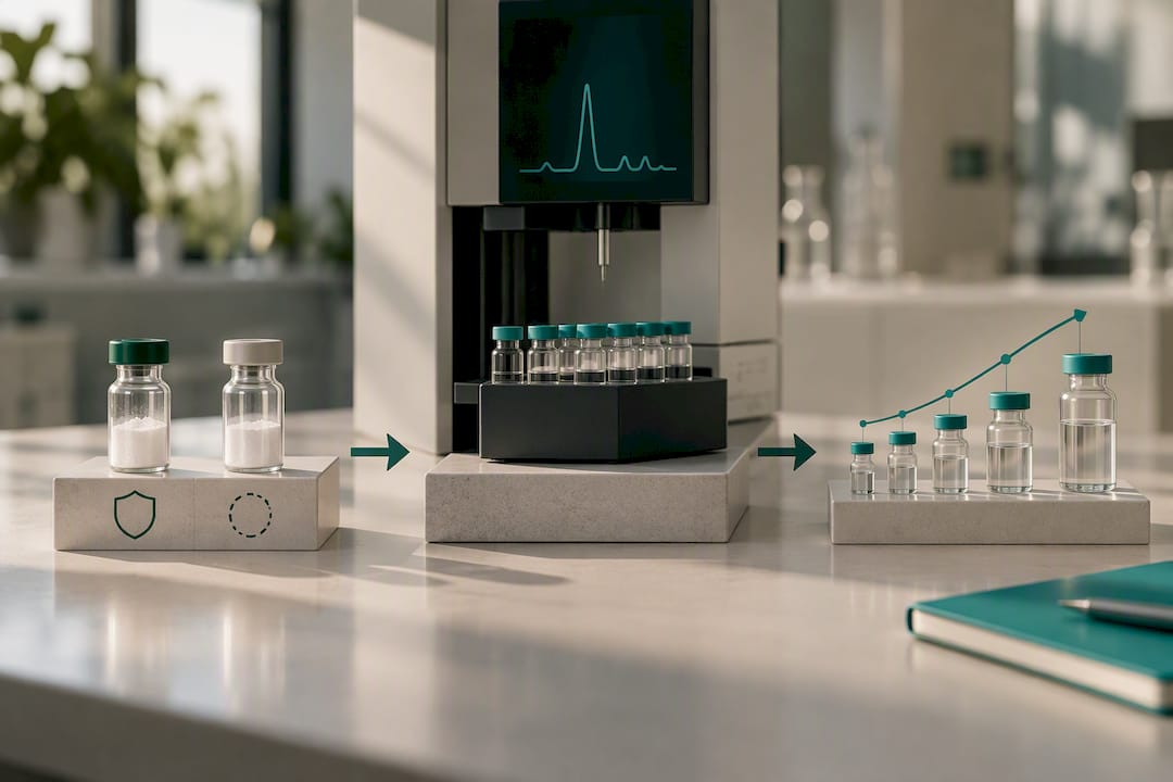
Peptastic Labs supplies KPV and related research peptides with batch-specific documentation built around the same verification standards this article recommends for any incoming lot. Every batch is independently tested and HPLC-verified to ≥99% purity, with Certificates of Analysis available on request. That documentation trail matters directly for the concerns raised above: a lab citing a specific batch ID and COA in its methods section makes replication by other groups far more credible than citing a peptide by name alone.
Quality control at this level, third-party verification layered on top of in-house checks, reduces one of the largest hidden sources of experimental variability in peptide research: unconfirmed identity or purity in the starting material. For studies where PepT1 uptake kinetics or NF-κB inhibition potency are the actual variables under test, starting with a peptide of confirmed purity removes a confound before the experiment begins.
- Request a Certificate of Analysis for any KPV lot before starting a new experimental series.
- Cross-reference incoming material against expected molecular identifiers for quality assurance.
- Review related peptide research on immune modulation for broader pathway context relevant to KPV's mechanism.
KPV Peptide Comparisons: How It Stacks Up Against Related Analogs
KPV's closest comparison point in the peptide research field is BPC-157, another short peptide studied heavily for gut and tissue-repair applications, though the two work through distinct mechanisms. Where KPV acts through PepT1-mediated uptake and direct NF-κB pathway inhibition, BPC-157's proposed mechanisms center more on angiogenesis and growth factor modulation in tissue repair contexts. Researchers exploring BPC-157 as a related research compound will find the two peptides address overlapping therapeutic areas, gut healing and inflammation, from different mechanistic angles, which makes them useful to study in parallel rather than as interchangeable options.
Within the melanocortin-derived peptide family, KPV's advantage is specificity. Full-length α-MSH and its longer analogs engage melanocortin receptors broadly, producing effects on pigmentation and appetite alongside any anti-inflammatory signal. KPV strips that complexity away, acting through PepT1 and NF-κB without the receptor-mediated side effects tied to the parent hormone. That makes it a cleaner experimental tool when the research question is specifically about NF-κB modulation rather than broader melanocortin biology.
Other short anti-inflammatory peptides studied in colitis models generally lack KPV's documented transporter-mediated entry mechanism, which is arguably KPV's most distinguishing feature in the literature. Most peptides rely on passive diffusion or receptor binding at the cell surface; KPV's active transport into the cell interior via PepT1 is a comparatively rare and well-characterized entry route among small research peptides.
Beyond NF-κB: Other Signaling Pathways KPV May Influence
NF-κB inhibition is KPV's best-documented mechanism, but it is unlikely to be the whole story. Inflammatory signaling networks are heavily cross-linked, and a peptide that blocks p65 nuclear import would be expected to produce downstream ripple effects across pathways that intersect with NF-κB activity.
Mitogen-activated protein kinase (MAPK) pathways, including p38 and JNK, run in parallel with NF-κB in most inflammatory cascades and often share upstream activators. Reduced NF-κB output can indirectly dampen MAPK-driven cytokine production even without KPV acting on MAPK components directly, which is an important distinction for interpreting any downstream cytokine data that shows broader suppression than NF-κB inhibition alone would predict.
Some researchers have also raised the question of whether KPV affects STAT (signal transducer and activator of transcription) signaling, given the tight functional overlap between NF-κB and STAT3 in chronic gut inflammation. This remains a hypothesis rather than an established finding in the current literature, and no completed mechanistic study has confirmed direct KPV activity on STAT pathways.
For experimental design, the practical takeaway is to avoid over-attributing every downstream effect to NF-κB inhibition alone. If your cytokine panel shows changes in mediators not classically downstream of NF-κB, a parallel pathway assay, a phospho-MAPK Western blot, for instance, can help distinguish direct KPV effects from secondary consequences of reduced NF-κB signaling. Treating NF-κB as the confirmed primary mechanism while staying open to secondary pathway involvement is the more defensible position given current evidence.
Measuring KPV Bioavailability: Why It's Harder Than It Sounds
Quantifying how much KPV actually reaches its target tissue intact is one of the more technically frustrating aspects of working with this peptide. Standard oral bioavailability studies rely on measuring parent compound concentration in plasma over time, but KPV's small size and rapid peptidase susceptibility make that approach unreliable without careful method development.
Mass spectrometry-based quantification is the standard tool, but it requires distinguishing intact KPV from its degradation fragments, which can be structurally similar enough to complicate detection specificity. Researchers working on the nanoparticle delivery approaches described earlier have had to develop tissue-specific extraction protocols just to get a clean read on colonic KPV concentration versus what remains in the upper GI tract.
Challenges that consistently come up in bioavailability work:
- Rapid enzymatic degradation in gastric and intestinal fluid makes timing of sample collection critical; a measurement taken even 30 minutes off can shift the apparent bioavailability substantially.
- PepT1-dependent uptake means tissue concentration of KPV does not scale linearly with dose in the way a passively absorbed drug would, complicating standard PK modeling assumptions.
- Distinguishing intracellular accumulated KPV from peptide still in the extracellular or luminal space requires cell fractionation steps that add time and variability to the protocol.
Given these challenges, most published studies report relative efficacy endpoints, DAI scores, cytokine levels, rather than absolute bioavailability figures, which is a reasonable workaround but leaves a real gap in the literature for anyone trying to model human dosing from mouse data.
Toxicity and Side Effects Observed in Preclinical Models
Preclinical toxicity data on KPV is limited but has not raised significant red flags in the studies conducted so far. The murine colitis studies that established KPV's anti-inflammatory efficacy did not report serious adverse events at the doses tested, and body weight recovery, one of the most sensitive general indicators of systemic toxicity in rodent studies, actually improved with treatment rather than declined.
That said, "no adverse events reported" in a handful of short-duration rodent studies is a narrow safety signal, not a completed toxicology profile. None of the published work has systematically evaluated chronic exposure, reproductive toxicity, or organ-specific effects across a range of doses in the way a full preclinical safety package for a drug candidate would require.
The MC1R-deficient mouse studies offer a useful side benefit here: because KPV worked without engaging melanocortin receptors, researchers can rule out receptor-mediated off-target effects tied to pigmentation or appetite regulation, two systems that full-length α-MSH analogs do affect. That narrows the plausible off-target risk profile somewhat, but it does not substitute for dedicated toxicology work.
Given this state of evidence, the responsible framing for KPV peptide dosage discussions in a research context is that preclinical data is reassuring within its limited scope, not that safety has been established. Any lab planning extended in vivo work should build in standard toxicology endpoints, liver and kidney function markers, complete blood counts, and histopathology of major organs, rather than relying on the absence of reported problems in prior published studies.
Next-Generation KPV: Stability and Activity Enhancement Research
The biggest engineering problem with KPV isn't its mechanism, it's how fast the unprotected peptide breaks down before reaching its target. That has pushed a meaningful share of current KPV peptide synthesis research toward modification strategies rather than the native sequence alone.
The hyaluronic acid-functionalized nanoparticle approach already discussed represents one branch of this work: encapsulation rather than modifying the peptide's own structure. A parallel branch focuses on direct chemical modification, exploring D-amino acid substitutions at one or more positions in the KPV sequence to resist protease cleavage while attempting to preserve the peptide's binding activity at its intracellular target. D-amino acid substitution is a well-established strategy in peptide chemistry generally, since many proteases are stereospecific and cannot cleave bonds involving D-configured residues.
Cyclization is another modification path researchers are exploring across short peptides broadly, and KPV is a plausible candidate given its size. A cyclized version could theoretically resist exopeptidase degradation, which typically attacks free N- or C-termini, though published cyclized-KPV data remains sparse.
PEGylation, attaching polyethylene glycol chains to extend circulation time, is a strategy more commonly applied to larger peptides and proteins, and its suitability for a tripeptide as small as KPV is less clear, since PEG conjugation risks masking the very structural features PepT1 recognizes for transport. Any modification strategy for KPV has to account for this tension: changes that improve stability must not simultaneously block the PepT1-mediated uptake that makes the native peptide effective in the first place.
What Would Actually Move KPV Research Forward
The field does not need more mouse colitis studies proving the same mechanism again. It needs a small, well-designed human Phase 1 safety and pharmacokinetic study, something that establishes basic tolerability and a plausible dose range in people. Standardized PepT1 expression panels across labs would also help, since right now every group measures uptake capacity slightly differently, which makes cross-study comparison harder than it should be.
Reproducibility depends on open methods and shared Certificates of Analysis becoming the norm rather than the exception. KPV is mechanistically one of the more interesting short peptides in the immunomodulatory peptide field, but interesting mechanism and clinical relevance are not the same claim, and the field should stop writing as though they are.
— Tintastic
Sourcing Research-Grade KPV for Your Next Study
Getting mechanistic KPV peptide research right depends on starting with material you can trust, and that means knowing exactly what purity level and batch history you are working with before the first assay runs. Peptastic Labs verifies every batch to ≥99% purity by HPLC, with independent testing and Certificates of Analysis available on request, giving you the documentation trail your methods section needs.
Research peptides sourced from verified vendors carry traceable batch documentation that can be cited directly when reviewers ask about material verification. The full product catalogue covers KPV alongside related research peptides across metabolic, immunology, and tissue-repair lines, with bulk options available for labs running larger study series. For background on experimental design and pathway context before you order, the research resources page has protocol guidance and peptide-specific write-ups worth reviewing first.
Record your batch ID, request the COA, and run your own confirmatory HPLC check on arrival. Visit the catalogue to place your order and get your next KPV lot into the lab with the documentation already in hand.
Primary Literature and Resources for Continued Reading
- PepT1-Mediated Tripeptide KPV Uptake Reduces Intestinal Inflammation — foundational mechanism and uptake kinetics study.
- Orally Targeted Delivery of Tripeptide KPV via Hyaluronic Acid-Functionalized Nanoparticles — nanoparticle delivery and mucosal healing data.
- PubMed entry summarizing KPV murine colitis findings — DSS/TNBS outcome data.
- PubMed Central (PMC) home — full-text archive for ongoing literature surveillance.
This article is general information, not a substitute for advice from a qualified doctor. Consult a qualified healthcare professional about your own circumstances before acting on anything here.
Sources
- PepT1-Mediated Tripeptide KPV Uptake Reduces Intestinal Inflammation
- Orally Targeted Delivery of Tripeptide KPV via Hyaluronic Acid-Functionalized Nanoparticles Efficiently Alleviates Ulcerative Colitis
- PubMed entry summarizing KPV murine colitis findings
- PubMed Central (PMC) home
FAQ
Does KPV peptide really work?
In cell culture and murine colitis models, KPV consistently reduces inflammatory markers through PepT1-mediated uptake and NF-κB inhibition, as shown in the foundational uptake study. Whether it "works" in humans is a different question, since no controlled human trials have tested clinical efficacy yet.
Who should not take KPV peptide?
KPV is a research compound without established human safety data, dosing, or regulatory approval for clinical use, so it isn't intended for use by anyone outside a supervised laboratory research setting. Pregnant or immunocompromised research subjects in animal models haven't been specifically characterized for KPV safety either, reinforcing that this remains preclinical-stage material.
How does KPV peptide compare to BPC-157?
KPV and BPC-157 both draw research interest for gut and inflammation applications but work through different mechanisms. KPV acts via PepT1 transport and direct NF-κB pathway inhibition, while BPC-157's proposed activity centers on angiogenesis and growth factor modulation, making them complementary rather than interchangeable research subjects.
How can I get KPV peptide?
Qualified researchers can source KPV through the Peptastic Labs catalogue, where each batch is HPLC-verified to ≥99% purity with a Certificate of Analysis available on request. Always confirm batch documentation before starting a new experimental series, regardless of vendor.