
For peptide identification and quantitation, LC–ESI–MS/MS bottom-up analysis is the most practical, widely used approach available to most labs. Success turns on three controls: clean, well-matched sample preparation, stable isotope labeled internal standards, and documented validation against a recognized framework. Get those three right and the rest of the method, from column choice to fragmentation strategy, becomes a matter of tuning rather than guesswork.
TL;DR:
- Ensuring sample preparation, stable isotope internal standards, and validation according to recognized frameworks is crucial for successful peptide mass spectrometry analysis.
- Choice of acquisition strategy (DDA, DIA, MRM, PRM) affects sensitivity, reproducibility, and suitability for discovery versus targeted quantitation.
- Digestion steps reduce sensitivity and introduce variability; skipping or minimizing digestion when analyzing synthetic peptides enhances detection limits.
- Using high-purity, HPLC-verified peptides with raw chromatogram data from trusted suppliers improves validation and troubleshooting efforts.
- Proper validation, including detailed documentation and resource controls, is essential to avoid relying on unconfirmed assumptions for peptide quantitation.
Table of Contents
- What Is the Standard Workflow for Mass Spectrometry Peptides Analysis?
- How Does Sample Preparation Affect Peptide Sensitivity?
- Which Chromatography and Mobile Phase Choices Work Best for Peptides?
- How Do Ionization Sources and Mass Analyzers Compare for Peptides?
- How Does Peptide Fragmentation Enable Sequence Identification?
- What Quantitation Strategy Should You Use for Peptides?
- What Are the Most Common Troubleshooting Issues in Peptide MS?
- How Do You Validate and Document Peptide Mass Spectrometry Assays?
- How Peptastic Labs Supports Peptide MS Research
- An Editorial Take on Getting Peptide MS Right the First Time
- Getting Documented, Research-Grade Peptides for Your MS Workflow
- Sources
- FAQ
What Is the Standard Workflow for Mass Spectrometry Peptides Analysis?
A bottom-up peptide mass spectrometry workflow moves through five stages: extraction, enzymatic or chemical preparation, liquid chromatography separation, ionization, and tandem mass spectrometry fragmentation followed by data interpretation. Each stage either preserves or destroys the information you need downstream, so the pipeline has to be planned backward from the question you're asking, not forward from whatever instrument happens to be free.
Bottom-up analysis, where proteins or synthetic peptides are digested or already exist as short chains before injection, dominates peptide LC-MS analysis because peptides fragment more predictably than intact proteins and produce cleaner b/y ion ladders. Intact (top-down) approaches have their place when you need to preserve a specific proteoform or confirm a full sequence without digestion artifacts, but they demand higher-resolution instrumentation and more careful calibration. For most synthetic peptide identity confirmation and quantitation work, bottom-up or direct-injection workflows on already-short peptides are the practical default.
The pipeline generally looks like this:
- Extraction and cleanup: pull the peptide of interest out of its matrix (plasma, cell lysate, or a reconstituted vial) while minimizing losses from adsorption.
- Preparation: digest with a protease if you're working from a protein, or quantify and dilute directly if you already have a synthetic peptide.
- Chromatographic separation: resolve peptides by hydrophobicity on a reversed-phase column before they reach the source.
- Ionization: convert separated peptides into gas-phase ions, almost always by electrospray in LC-coupled setups.
- Fragmentation and detection: generate MS/MS spectra and match them against a database or interpret them de novo.
Acquisition strategy shapes everything downstream. Data-dependent acquisition (DDA) selects the most abundant ions in real time for fragmentation, which works well for broad discovery work but can miss lower-abundance species run to run. Data-independent acquisition (DIA) fragments everything in defined mass windows regardless of intensity, trading some spectral clarity for far better reproducibility across samples, which matters when you're comparing cohorts over weeks or months. Targeted approaches like multiple reaction monitoring (MRM) or parallel reaction monitoring (PRM) skip discovery altogether and watch only the transitions you already know matter, delivering the lowest LLOQs and highest reproducibility for quantitation work once a target list is locked in. Foundational peptide and protein MS reviews describe these workflow variations in more depth, and they're worth keeping on hand when you're designing a new assay from scratch.
How Does Sample Preparation Affect Peptide Sensitivity?
Digestion is not free. A study quantifying salmon calcitonin found an LLOQ of 10 pg/mL when the intact peptide was measured directly, but that LLOQ rose to 50 pg/mL once the sample was tryptically digested and quantitated through a signature peptide. Digestion introduces missed cleavages, incomplete reactions, and a flood of unrelated peptide fragments competing for ionization, and all of that complexity eats into your limit of quantitation.
That does not mean digestion is wrong. It means the decision has to be deliberate. Trypsin remains the default protease because it cuts predictably after lysine and arginine, generating peptides in the ideal 700 to 3,000 Da range for most triple quadrupole and Orbitrap workflows. Use it when you're working from intact proteins and need a signature peptide as a proxy. Skip digestion entirely, or minimize sample handling, when you're already working with a synthetic peptide and identity confirmation or direct quantitation is the goal. Every unnecessary step between vial and injection port is another chance to lose signal.
Practical cleanup matters just as much as the enzymatic step:
- Solid-phase extraction (SPE) or desalting removes salts and detergents that suppress ionization before the sample ever reaches the column.
- Basic peptides (high arginine or lysine content) tend to stick to stainless steel surfaces and tubing, so low-adsorption plasticware and acidified buffers reduce losses.
- Peptide quantification before injection using a microvolume UV/visible spectrophotometer, rather than assuming a nominal concentration, catches degradation and concentration drift that would otherwise masquerade as instrument variability.
One study using a microfluidic UV/visible device found that direct peptide quantification in the MS-loading solvent, using only 2 microliters of sample, meaningfully improved consistency between identification and quantitation runs. The same research recommended a practical injected peptide amount for many LC–MS/MS setups around a few micrograms as a starting point rather than a hard rule, since column capacity and source design both shift the optimum.
Pro Tip: Quantify your peptide stock immediately before dilution for injection, not at the time the vial was reconstituted. A stock sitting at 4°C for two weeks can lose measurable mass to adsorption on the vial wall, and you'll never see it if you trust the label concentration instead of measuring fresh.
Controlling injected mass isn't a cosmetic detail. Overloading a column degrades peak shape and promotes ion suppression from co-eluting species; underloading pushes you below your assay's working range. Both failure modes look identical on a chromatogram until you trace them back to load.
Which Chromatography and Mobile Phase Choices Work Best for Peptides?
Column and mobile phase selection for peptide LC-MS analysis comes down to one recurring tension: what separates peptides best on a UV detector often suppresses their ionization in the mass spectrometer. Trifluoroacetic acid (TFA) has been a chromatography workhorse for decades because it produces sharp, symmetric peptide peaks through strong ion-pairing, but TFA is a well-documented ion suppressor in electrospray sources, sometimes cutting signal intensity dramatically compared to formic acid.
Formic acid (FA) is the standard alternative for MS-facing methods because it doesn't compete with peptide ions for charge in the source. The trade-off has traditionally been peak shape: FA alone often gives broader, less resolved peaks for peptides, especially basic ones, than TFA-based methods deliver. Specialized peptide-optimized stationary phases, including charged-surface superficially porous particles, narrow that gap by improving peak shape under FA conditions well enough to make FA-only methods viable for both impurity profiling and identity confirmation work.
Column choices worth knowing:
- 120 Å superficially porous particles balance peptide diffusion and resolution better than fully porous particles for peptides in the 500 to 5,000 Da range.
- Charged-surface columns reduce basic peptide tailing without resorting to ion-pairing reagents that hurt MS sensitivity.
- Wide-pore C18 phases remain the default starting point for general peptide separations before you troubleshoot toward something specialized.
Sample composition and injection solvent quietly determine whether any of this works. A peptide dissolved in an organic-heavy solvent that doesn't match your mobile phase A will often elute as a distorted peak, or fail to focus on the column head at all. Match the injection solvent's organic content to your starting mobile phase composition whenever possible, and don't assume a method that worked for one peptide will behave identically for a structurally different one.
Method transfer between LC/UV development and LC–MS deployment is where a lot of labs lose time. A TFA-based UV method that looked clean on development equipment can produce a flattened, suppressed signal the moment it moves to a mass spectrometer. Building the method on FA-compatible peptide columns from the start, even during early UV-only development, avoids re-optimizing the separation twice.
How Do Ionization Sources and Mass Analyzers Compare for Peptides?
Electrospray ionization (ESI) is the default choice for peptide LC-MS analysis because it couples directly with liquid chromatography and produces multiply charged ions that extend the effective mass range of almost any analyzer. MALDI, by contrast, ionizes from a dried crystalline matrix rather than a liquid stream, which makes it faster for high-throughput spot-based screening but poorly suited to online LC separation. Choose MALDI when you're profiling many discrete samples without chromatography attached; choose ESI whenever separation matters.
Analyzer choice depends on the question. Orbitrap and time-of-flight (TOF) instruments deliver the high-resolution, accurate mass (HRMS) data that discovery proteomics and de novo sequencing depend on, resolving isobaric species and confirming elemental composition from mass accuracy alone. Triple quadrupole instruments sacrifice that resolution for sensitivity and speed in targeted work, and they remain the standard for MRM-based quantitation where you already know your target transitions and just need to measure them reliably across hundreds of samples.
Flow rate is where a lot of labs make assumptions that cost them sensitivity. Nanoflow LC, running at a few hundred nanoliters per minute, maximizes the proportion of peptide ions that form in the gas phase relative to solvent, and it remains the sensitivity benchmark for discovery proteomics. But nanoflow is fragile, prone to clogging, and slow to equilibrate between runs, which makes it a poor fit for high-throughput quantitative labs running dozens of samples a day.
Advanced ion source designs close some of that gap. Jet Stream and ion funnel (iFunnel) technologies focus ions more efficiently before they enter the analyzer, letting conventional-flow chromatography, running at hundreds of microliters per minute instead of nanoliters, approach nanoflow-level sensitivity for many peptide panels. One application note using an Agilent triple quadrupole with Jet Stream ionization reported LLOQs as low as 5 attomoles per microliter for well-responding peptides paired with stable isotope internal standards. That kind of performance means you don't always have to sacrifice throughput to hit demanding sensitivity targets, though the achievable LLOQ still depends heavily on the specific peptide's ionization efficiency and matrix background.
How Does Peptide Fragmentation Enable Sequence Identification?
Peptide fragmentation mass spectrometry works by breaking the peptide backbone at the amide bond during MS/MS, generating two complementary ion series: b-ions, which retain the N-terminal fragment, and y-ions, which retain the C-terminal fragment. Reading the mass differences between consecutive ions in either series reveals the amino acid sequence one residue at a time, which is the entire basis for peptide identification mass spectrometry.

The fragmentation method you choose changes what that spectrum looks like. Collision-induced dissociation (CID) and higher-energy collisional dissociation (HCD) both fragment predominantly at the amide bond to produce clean b/y ladders, with HCD generally giving better fragment ion statistics on modern Orbitrap instruments. Electron transfer dissociation (ETD) fragments differently, preserving labile post-translational modifications like phosphorylation that CID and HCD often strip away before you can localize them. If PTM site localization matters for your peptide, ETD (or a hybrid method combining it with HCD) usually outperforms CID alone.
Database searching against known sequences is sufficient for the overwhelming majority of peptide identification mass spectrometry work, especially for synthetic peptides where the expected sequence is already known and the task is confirmation rather than discovery. De novo sequencing, which reconstructs sequence directly from spectral mass differences without a reference database, earns its place when you're characterizing an unknown impurity, a novel modification, or a degradation product with no matching database entry. Algorithm quality here has improved substantially. A tool called Spectralis demonstrated markedly better sensitivity and precision on benchmark spectra compared to earlier de novo methods, narrowing the reliability gap that used to make de novo a last resort.
Whichever route you take, false discovery rate (FDR) control is non-negotiable for defensible identification. Standard practice reports peptide-level FDR at 1%, typically through target-decoy database searching, and any identification claim without a stated FDR threshold should be treated with suspicion.
What Quantitation Strategy Should You Use for Peptides?
Targeted MRM or PRM quantitation with stable isotope labeled (SIL) internal standards is the most reliable approach for peptide quantitation LC-MS work where absolute concentration matters. Label-free quantitation, comparing raw peak areas or spectral counts across runs without a labeled standard, works reasonably well for relative comparisons within a single discovery experiment but accumulates too much run-to-run variability for absolute quantitation across batches or labs.
SIL peptides, chemically identical to your analyte but incorporating heavy carbon-13 or nitrogen-15 labeled amino acids, co-elute with the native peptide and correct for matrix effects, ionization efficiency drift, and extraction losses because they experience nearly identical chemistry throughout the workflow. When a true SIL standard isn't available or affordable, a structurally similar peptide analog can serve as an internal standard, though it corrects for fewer sources of variability and should be validated carefully before you trust it.
LLOQ in any peptide assay is driven by three interacting factors:
- Matrix complexity: plasma and serum introduce far more ion suppression than a clean buffer system.
- Digestion strategy: as covered earlier, digesting a sample can push LLOQ from 10 pg/mL up to 50 pg/mL for the same analyte depending on signature peptide choice.
- Transition selection: choosing MRM transitions with minimal interference from co-eluting isobaric species tightens the achievable LLOQ substantially.
With the right instrumentation and standards, some peptide panels achieve LLOQs in the low attomole-per-microliter range, but that ceiling depends entirely on ionization efficiency for the specific peptide and how clean the matrix background is, not on the instrument alone.
A minimum validation checklist, aligned with CLSI C64 principles, should cover: a clearly defined measurand (the exact peptide form and any modification state you're measuring), documented calibrator and internal standard sourcing, pre-specified accuracy and precision acceptance criteria, and a formal validation run before the method goes into routine use. Skipping the measurand definition step is the single most common source of downstream validation disputes, because "quantifying the peptide" means something different depending on whether you're measuring free peptide, total peptide, or a specific proteoform.
What Are the Most Common Troubleshooting Issues in Peptide MS?
Low signal, distorted peaks, and irreproducible quantitation almost always trace back to one of four root causes. Work through them in this order before you start changing instrument parameters:
- Check for ion suppression first. Co-eluting matrix components, residual detergents, or TFA in your mobile phase can silently crush signal without any obvious chromatographic clue. Run a post-column infusion test or compare signal in neat solvent versus matrix to confirm.
- Evaluate peak shape for basic peptides. Tailing or splitting peaks on arginine or lysine-rich peptides usually mean you need either a mild ion-pairing compromise or a switch to a charged-surface column designed for basic analytes.
- Rule out carryover and adsorption. Run a blank injection immediately after your highest-concentration standard; if you see a ghost peak, autosampler carryover is diluting your quantitation accuracy in every subsequent run.
- Reoptimize transition selection. If two peptides in your panel share a similar precursor or product ion mass, interference will inflate quantitation at low concentrations specifically, which is exactly where you can least afford it.
Injection solvent composition deserves its own line item. A mismatch between injection solvent and mobile phase A causes peptides to elute as broad, misshapen peaks or, in bad cases, to pass straight through the column without retention at all. When in doubt, dilute your injection solvent toward your starting mobile phase composition rather than assuming a stronger solvent will "just work."
Pro Tip: Keep a rotating log of blank injections between sample batches, not just at the start of a run. Carryover often builds gradually across a plate and only becomes visible three or four samples in, well past the point where a single start-of-run blank would have caught it.
How Do You Validate and Document Peptide Mass Spectrometry Assays?
CLSI C64 lays out a four-stage framework for developing and validating quantitative peptide and protein assays by LC–ESI–MS/MS: define the measurand precisely, select appropriate calibrators and internal standards, set numerical performance criteria for accuracy and precision, and execute a formal validation run against those criteria. Following that sequence, rather than validating opportunistically after a method is already in routine use, catches measurand ambiguity and internal standard mismatches before they become buried assumptions in a dataset you've already published.
HRMS paired with MS/MS confirmation is the standard combination for impurity profiling and identity confirmation of synthetic peptides. High-resolution accurate mass narrows candidate elemental compositions for an unknown impurity peak, while fragmentation confirms sequence and localizes any modification. A typical endpoint for identity confirmation combines a mass accuracy within a few parts per million of the theoretical value with a fragmentation pattern matching the expected sequence.
What should you request from a peptide supplier before building validation records around their material? At minimum:
- A Certificate of Analysis documenting purity by HPLC and confirming identity by mass spectrometry.
- Raw chromatograms, not just summary purity percentages, so you can independently assess peak shape and detect co-eluting impurities the summary number might mask.
- Batch-specific data, since purity and impurity profiles can shift meaningfully between production lots even for the same nominal peptide.
Building these documents into your own validation records from the outset, rather than treating them as separate paperwork, gives you a defensible chain of evidence if a result is ever questioned later.
How Peptastic Labs Supports Peptide MS Research
Peptastic Labs independently tests its research-grade peptide catalog to HPLC-verified purity of at least 99%, with Certificates of Analysis available on request for every batch. That documentation isn't decorative. A CoA functions as a QC checkpoint you can drop directly into your own validation record, confirming the identity and purity baseline your MS assay is measuring against before you've run a single sample.
Raw chromatograms matter more than summary purity figures for exactly the reason covered in the validation section above: a single percentage can hide a co-eluting impurity that only becomes visible when you look at the trace itself. Where those chromatograms are available, they give researchers a reference point to compare against their own LC–MS/UV output and catch discrepancies before they become published errors.
Practical ways to use these artifacts in your own workflow:
- Cross-check CoA purity against your own HRMS identity confirmation before treating a peptide as validated in your assay.
- Use raw chromatogram peak shape as a baseline when troubleshooting your own column and mobile phase performance.
- Reference batch documentation when a result looks anomalous, since a batch-specific impurity shift is often the simplest explanation.
Peptastic Labs also maintains educational research resources covering third-party testing practices and purity standards, useful background when you're deciding how much independent verification a given research application warrants. For labs building out orthogonal confirmation methods, such as immunoenrichment ahead of MS, reagent selection guides like Assay Genie's polyclonal versus monoclonal comparison cover a decision point that shows up often in front-end sample prep.
An Editorial Take on Getting Peptide MS Right the First Time
Most peptide MS methods fail not at the mass spectrometer but at the bench, hours before the sample ever reaches the source. The conventional wisdom treats digestion as an automatic first step and internal standards as a nice-to-have you'll add if time and budget allow. Both habits are backwards. Digestion should be a deliberate choice weighed against a documented sensitivity cost, and SIL standards should be budgeted into a method from the design stage, not bolted on when a validation run fails.
If there's one action plan worth taking away from all of this, it's sequential: pick your acquisition mode based on the actual question (discovery calls for DDA or DIA, targeted quantitation calls for MRM or PRM), validate your transitions before you trust a single number, use SIL peptides wherever the budget allows, quantify your peptide stock immediately before each run rather than trusting a label, and collect a Certificate of Analysis with raw chromatograms from your peptide source before you build a validation record around it. Skip any one of those five steps and you're not running a validated assay. You're running an educated guess with good instrumentation attached.
— Tintastic
Getting Documented, Research-Grade Peptides for Your MS Workflow
Some peptide sources provide a Certificate of Analysis and raw chromatogram data, which can be used directly in validation records instead of relying solely on purity claims. Every compound in the catalog is HPLC-verified to at least 99% purity and independently tested before it ships, which means the identity confirmation step covered earlier in this guide starts from documented ground rather than a guess.
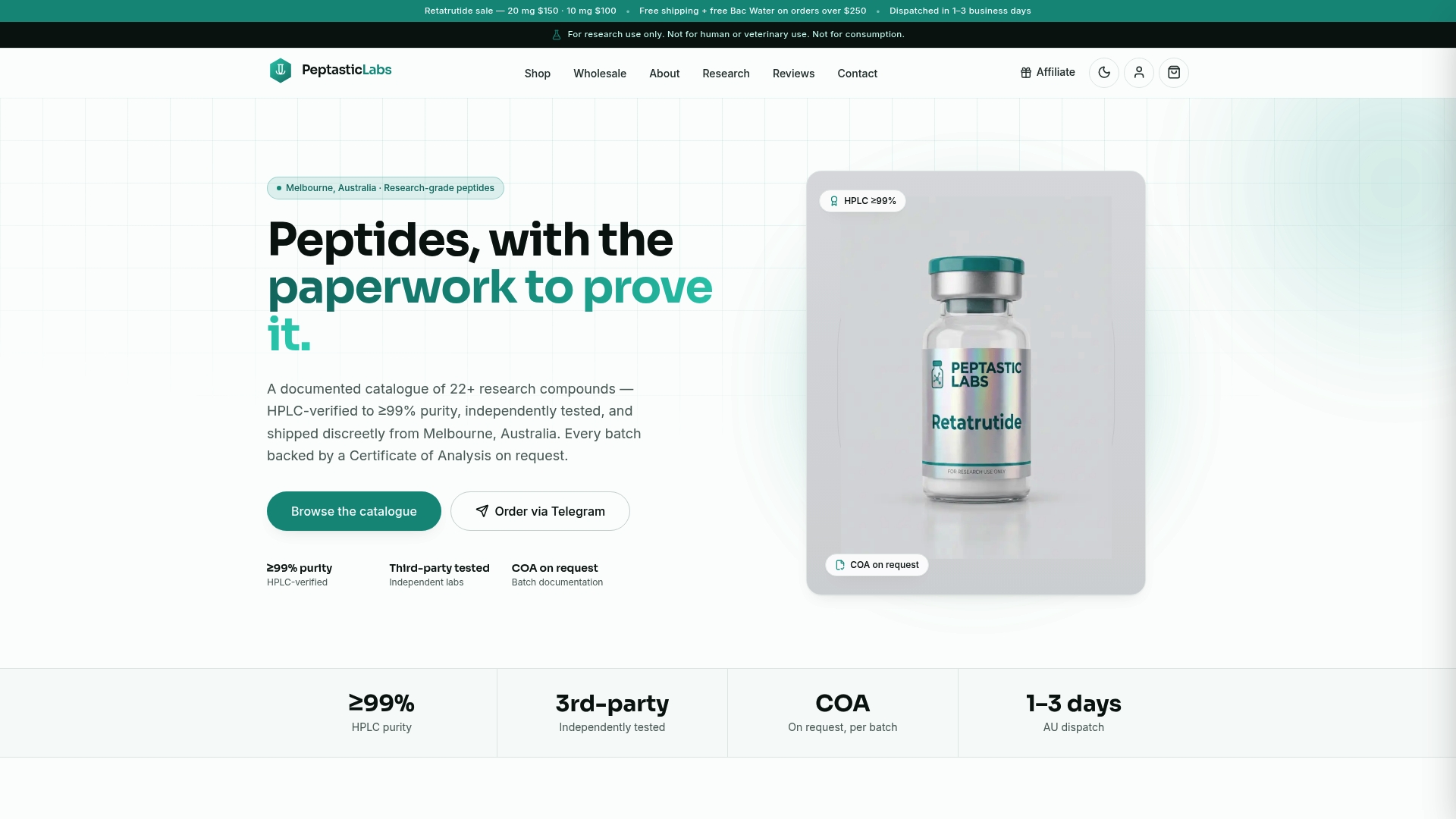
That documentation matters most at the exact moment your method validation runs into a discrepancy, when you need to know whether an unexpected peak came from your chromatography or from the peptide itself. Full batch traceability across the catalog lets you check that before it costs you a data point. Compounds like MOTS-c ship with the same testing rigor as the rest of the line, so switching between research applications doesn't mean switching your confidence level in the material.
If you're setting up a new LC–MS/MS assay or troubleshooting an existing one, start by requesting a Certificate of Analysis for the specific batch you're working with through the Peptastic Labs catalog. Options for bulk and wholesale purchases may be available for labs running larger validation studies across multiple peptide targets.
FAQ
Is 99% Purity Good for Research Peptides?
Purity alone doesn't guarantee assay success, though; matrix effects and digestion choices still shape what LLOQ you can actually achieve downstream.
How Can You Tell if a Peptide Has Degraded?
Degradation typically shows up as unexpected extra peaks on an HPLC or LC–MS trace, a shift in retention time from the reference chromatogram, or a mass shift in HRMS consistent with oxidation, deamidation, or cleavage. Comparing a fresh chromatogram against the original Certificate of Analysis trace is the fastest way to catch it.
How Do You Identify a Peptide Bond in Mass Spectrometry?
A peptide bond fragments during MS/MS to generate the complementary b-ion and y-ion series that reveal sequence, with the mass difference between adjacent ions in either series corresponding to a specific amino acid residue. Fragmentation methods like HCD, CID, or ETD each break that bond with slightly different efficiency and modification retention.
What Fragmentation Method Preserves Post-Translational Modifications Best?
Electron transfer dissociation (ETD) preserves labile modifications like phosphorylation more reliably than CID or HCD, which often strip these groups off before the fragment ions can be measured. Many labs combine ETD with HCD in a hybrid method to get both PTM retention and strong overall fragment ion coverage.
When Should You Use De Novo Sequencing Instead of Database Search?
Database searching is sufficient when you already know the expected peptide sequence, which covers most synthetic peptide identity confirmation work. De novo sequencing earns its place when you're characterizing an unknown impurity or novel modification with no matching database entry, and modern algorithms have narrowed the reliability gap with database methods considerably.