
Peptides function as direct biochemical signals in collagen production, binding to receptors on dermal fibroblasts and triggering upregulation of COL1A1, ELN, and VCAN gene expression. Research published in Frontiers in Medicine confirmed that collagen peptides significantly increased COL1A1, ELN, and VCAN gene expression in human dermal fibroblasts (p < 0.005), with confocal microscopy showing measurable increases in collagen synthesis after 24 hours of treatment. The primary cellular targets are dermal fibroblasts, the most abundant collagen-producing cells in connective tissue.
Key peptide types involved in collagen regulation include:
- Dipeptides and tripeptides (e.g., Pro-Hyp): Resist enzymatic degradation, reach dermal tissue intact, and directly stimulate fibroblast activity
- Signal peptides (e.g., Matrixyl/palmitoyl pentapeptide-4): Mimic collagen breakdown fragments, prompting fibroblasts to upregulate collagen I, III, and IV synthesis
- Carrier peptides (e.g., GHK-Cu): Transport copper into tissue, supporting collagen cross-linking and elastin maturation
- Enzyme-inhibiting peptides: Block MMP-1 and MMP-3, reducing collagen and elastin degradation within the extracellular matrix (ECM)
Both synthesis stimulation and degradation inhibition contribute to net collagen accumulation, making peptide class selection critical for research design.
Table of Contents
- How peptides activate collagen synthesis pathways in fibroblasts
- What recent studies reveal about collagen peptide supplementation
- Regulatory perspectives and realistic expectations for collagen peptide research
- Quality standards for research-grade peptides in collagen studies
- Endogenous versus exogenous peptides in collagen regulation
- Peptasticlabs: research-grade peptides for collagen biology
- Key Takeaways
How peptides activate collagen synthesis pathways in fibroblasts
Peptide bioavailability depends heavily on molecular size and amino acid composition. Hydroxyproline-rich di- and tripeptides like Pro-Hyp resist degradation during gastrointestinal transit and reach dermal fibroblasts at concentrations sufficient to trigger signaling cascades. Larger collagen fragments are hydrolyzed to free amino acids before absorption, distributing systemically rather than targeting skin directly.
Once at the fibroblast, peptides activate several converging pathways:
- TGF-β pathway: Signal peptides like Matrixyl activate transforming growth factor-beta signaling, directly instructing fibroblasts to synthesize collagen I, III, and IV
- PI3K-Akt, MAPK, and mTOR pathways: Daily 15 g collagen peptide combined with resistance training activates these anabolic pathways, supporting tendon and musculoskeletal collagen remodeling
- MMP inhibition: Collagen peptides significantly decrease MMP-1 and MMP-3 levels in fibroblast cultures, preserving ECM integrity; antioxidant co-treatment amplifies this effect
Vitamin C is a non-negotiable cofactor in this process. Hydroxylation of proline and lysine residues, catalyzed by prolyl and lysyl hydroxylases, requires ascorbate as an electron donor. Without adequate vitamin C, newly synthesized procollagen chains cannot form stable triple helices, and collagen maturation stalls regardless of peptide signaling activity.
Pro Tip: When designing collagen synthesis assays, verify that your peptide source provides documented bioactive sequences, specifically hydroxyproline-containing species. Sequence integrity directly determines whether a peptide activates the intended receptor-mediated pathway or produces non-specific background signal.
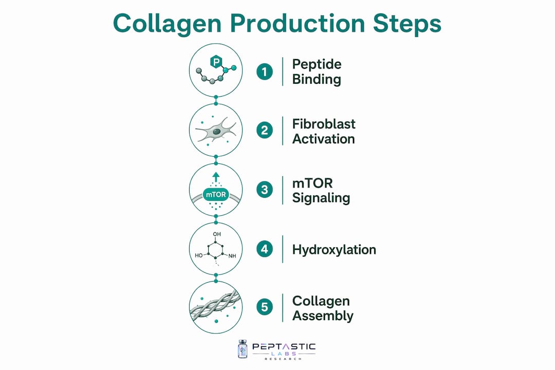
For researchers studying mTOR pathway activation in collagen contexts, confirming the precise peptide sequence and purity before experimental use prevents confounded results.
What recent studies reveal about collagen peptide supplementation
The 2024–2025 literature has substantially clarified both the efficacy and the mechanisms behind collagen peptide supplementation in skin and musculoskeletal models.
In a skin collagen-deficient mouse model, collagen tripeptide supplementation increased skin collagen content significantly (https://pubs.rsc.org/fo/article/16/13/5326-5344/893105) compared to controls. Transcriptomic analysis attributed this to TGF-β pathway activation and suppression of inflammation-related collagen degradation. Gut microbiota analysis showed increased alpha diversity (Shannon index) and elevated abundance of SCFA-producing bacteria, Lachnoclostridium and Roseburia, with Pro-Hyp delivery to the hindgut identified as the mechanistic link between supplementation and systemic TGF-β activation.
119.95% increase in skin collagen content was recorded in a collagen-deficient mouse model following collagen tripeptide supplementation, with TGF-β pathway activation confirmed by transcriptomic analysis.
In human musculoskeletal research, long-term collagen peptide supplementation produced statistically significant improvements in fat-free mass and tendon morphology (p < 0.01). The combination of 15 g daily collagen peptide with resistance training specifically boosted tendon cross-sectional area and activated PI3K-Akt, MAPK, and mTOR signaling. For researchers studying musculoskeletal collagen remodeling, these pathway-level findings provide a mechanistic basis for designing intervention protocols.
Skin-specific outcomes across clinical studies include measurable improvements in hydration, elasticity, and collagen density. The Frontiers in Medicine in vitro data showing COL1A1 upregulation at both 0.01% and 1% peptide concentrations after 24 hours establishes a dose-response relationship relevant to topical and injectable research models.

Regulatory perspectives and realistic expectations for collagen peptide research
Cleveland Clinic guidance classifies collagen peptides as "possibly effective" for skin hydration, elasticity, and knee osteoarthritis pain relief, while noting the absence of large-scale randomized controlled trials. This framing positions collagen peptides as maintenance-supporting compounds rather than therapeutics with definitive clinical proof.
A critical distribution limitation applies to oral supplementation: ingested collagen peptides are digested to amino acids that distribute systemically according to physiological demand, not preferentially to skin or joints. Researchers designing oral supplementation studies should account for this in outcome measurement and tissue sampling protocols.
Factors that directly affect research validity and reproducibility:
- Peptide molecular weight and sequence: Hydroxyproline-containing tripeptides show superior absorption versus high-molecular-weight collagen; sequence verification is required
- Dose and duration: Most positive human data involves multi-week protocols; single-dose or short-term designs may not capture ECM remodeling effects
- Concomitant variables: Physical training, vitamin C status, and antioxidant co-treatment all modulate outcomes
- Model selection: Mouse and rat models show larger effect sizes than human trials; extrapolation requires caution
- Outcome measurement: Skin biopsy with histological confirmation, as used in Matrixyl RCT data, provides stronger evidence than surface measurements alone
More large-scale, placebo-controlled human trials are needed before collagen peptide supplementation can be characterized as therapeutically definitive for any indication.
Quality standards for research-grade peptides in collagen studies
Reproducible collagen research depends on peptide purity and characterization that meet or exceed established benchmarks. Contaminants, degradation products, and sequence variants introduce confounding variables that invalidate mechanistic conclusions.
Research-grade quality markers for collagen-related peptides:
- Purity: ≥99% verified by HPLC; this threshold distinguishes research-grade material from commercial supplement-grade product
- Sequence integrity: Confirmed by mass spectrometry; critical for hydroxyproline-containing species where post-translational modification status affects bioactivity
- Homogeneity: Single-species preparations; mixed peptide pools obscure receptor-binding specificity in fibroblast assays
- Contaminant absence: Endotoxin testing required for cell culture applications; lipopolysaccharide contamination activates inflammatory pathways that confound collagen synthesis readouts
- Batch documentation: Certificate of Analysis (CoA) per lot, with HPLC chromatograms and mass spec data available on request
Storage and handling also affect experimental validity. Lyophilized peptides should be stored at -20°C or below, reconstituted in appropriate solvent immediately before use, and not subjected to repeated freeze-thaw cycles. Batch-to-batch consistency, confirmed by CoA comparison across lots, is required for longitudinal studies.
Researchers sourcing peptides for collagen biology work should consult a research-grade peptide guide to verify supplier documentation standards before committing to a source. The peptide types and uses resource from Peptasticlabs provides classification detail relevant to selecting the correct peptide class for specific collagen pathway targets.
Endogenous versus exogenous peptides in collagen regulation
Endogenous collagen-related peptides arise from enzymatic degradation of existing collagen fibrils. Fragments such as GHK (glycyl-L-histidyl-L-lysine) occur naturally in human plasma, urine, and saliva, and function as feedback signals that upregulate collagen synthesis when tissue degradation is detected. This endogenous signaling loop is concentration-dependent; GHK levels decline with age, correlating with reduced fibroblast activity and slower ECM repair.
Exogenous peptides introduced via supplementation or topical application work through the same receptor-mediated pathways but at controlled, experimentally defined concentrations. The key distinction is specificity: endogenous peptides are generated stochastically from degradation events across multiple tissue compartments, while exogenous peptides can be delivered as defined sequences targeting specific pathways. GHK-Cu, for example, modulates expression of over 4,000 human genes and activates copper-dependent lysyl oxidase, the enzyme responsible for collagen cross-linking. Endogenous GHK achieves this only when degradation-derived concentrations are sufficient, which declines with age.
For research design, this distinction matters. Studies using exogenous peptides can isolate pathway-specific effects that endogenous signaling cannot, because the endogenous pool is always a mixture of fragments from multiple collagen types and degradation stages. Exogenous peptides with verified sequences allow receptor-binding studies, dose-response characterization, and pathway inhibition experiments that are not feasible with endogenous fragment mixtures. The tissue repair signaling context for exogenous peptides is well-documented and directly applicable to collagen biology experimental design.
Peptasticlabs: research-grade peptides for collagen biology
Researchers working on collagen synthesis, ECM remodeling, or fibroblast signaling need a peptide source that eliminates purity uncertainty from the experimental equation. Peptasticlabs supplies over 22 independently tested compounds, each verified to ≥99% purity via HPLC, with Certificates of Analysis available per batch.

For collagen-focused research, the catalog includes BPC-157 for tissue repair and ECM studies, TB-500 for wound healing and collagen remodeling protocols, and the Glow compound targeting skin collagen and extracellular matrix restoration. Third-party verification and batch documentation are standard, not optional. Researchers can request CoA documentation before committing to a lot, supporting experimental reproducibility across multi-phase studies. Review the full catalog at Peptasticlabs to match compound specifications to your collagen research protocol.
Key Takeaways
Peptides drive collagen production by activating fibroblast signaling pathways, with hydroxyproline-rich tripeptides showing the strongest bioavailability and a documented 119.95% increase in skin collagen content in controlled models.
| Point | Details |
|---|---|
| Peptides as signaling molecules | Collagen peptides upregulate COL1A1, ELN, and VCAN gene expression in dermal fibroblasts (p < 0.005). |
| Key mechanistic pathways | TGF-β, PI3K-Akt, MAPK, and mTOR pathways mediate fibroblast activation and ECM remodeling. |
| Supplementation evidence | Collagen tripeptides increased skin collagen content by 119.95% in a mouse model; human data shows significant tendon morphology improvements (p < 0.01). |
| Regulatory framing | Cleveland Clinic classifies collagen peptides as "possibly effective"; large-scale RCTs remain limited. |
| Peptasticlabs quality standard | Over 22 compounds verified to ≥99% purity via HPLC, with batch-specific Certificates of Analysis on request. |